
The ongoing quest for faster, safer, and more comprehensive diagnostic imaging has driven a revolution in medical technology, pushing beyond the well-established yet limited capabilities of conventional methods. A novel, noninvasive 3D imaging system developed through a collaborative effort between the Keck School of Medicine of USC and the California Institute of Technology represents a significant leap forward in this pursuit. This review explores the evolution of this hybrid photoacoustic ultrasound technology, detailing its core features, performance, and transformative impact across various clinical applications. The purpose is to provide a thorough understanding of the system’s current capabilities and its trajectory toward becoming a mainstream diagnostic tool.
Foundational Principles of Hybrid Imaging
The emergence of hybrid photoacoustic ultrasound technology addresses a critical gap in medical diagnostics, and its core principle lies in the synergistic combination of two distinct imaging modalities—ultrasound and photoacoustics—to generate a single, comprehensive dataset. This approach is designed to overcome the persistent limitations of standard imaging tools like computed tomography (CT) and magnetic resonance imaging (MRI), which often involve trade-offs between cost, radiation exposure, scan time, and image resolution. By integrating complementary information sources, the hybrid system provides insights that are greater than the sum of its parts.
This technology has emerged in a context where clinical needs are rapidly evolving. While conventional methods remain invaluable, their constraints can hinder timely and accessible diagnostics. For instance, CT scans involve ionizing radiation, and MRI is expensive and time-consuming, making both unsuitable for frequent monitoring or use in resource-limited settings. The hybrid photoacoustic ultrasound platform offers a compelling alternative by providing rapid, radiation-free imaging at a potentially lower cost, thereby promising to enhance diagnostic workflows and expand access to advanced medical imaging.
A Deep Dive into the RUS-PAT System Architecture
Capturing Anatomy with Rotational Ultrasound Tomography
The architectural foundation of the hybrid system’s anatomical imaging capability is Rotational Ultrasound Tomography (RUST). This component represents a significant evolution from traditional handheld ultrasound. Instead of a single probe generating a two-dimensional image slice, RUST utilizes a sophisticated arc of detectors that direct sound waves into the body from multiple angles. This array-based approach allows for a much wider field of data acquisition during a single rotational scan.
By capturing the returning sound echoes from this comprehensive sweep, the system computationally reconstructs a full 3D volumetric image of the body’s soft tissue structures. This process yields a detailed anatomical map, providing the essential structural context needed for accurate diagnosis. The result is a high-fidelity rendering of tissues and organs that serves as the framework upon which functional data is overlaid.
Illuminating Vasculature with Photoacoustic Tomography
Working in tandem with RUST is the Photoacoustic Tomography (PAT) component, which provides a detailed map of the body’s vasculature. The PAT mechanism begins with the emission of a harmless, short-pulsed laser beam directed at the target tissue. The light energy from this laser is selectively absorbed by hemoglobin, the oxygen-carrying protein abundant in red blood cells. This targeted energy absorption creates a unique and highly useful physical reaction.
Upon absorbing the laser pulse, hemoglobin molecules undergo a minuscule but rapid thermal expansion and contraction. This movement generates high-frequency sound waves that propagate outward through the tissue. These light-induced acoustic signals are then detected by the same arc of transducers used for the RUST imaging. By analyzing these signals, the system constructs a precise, high-resolution 3D map of the blood vessels, effectively visualizing the body’s circulatory network.
Creating a Unified View Through Data Fusion
The true innovation of the RUS-PAT system lies in its ability to synergistically integrate the datasets from both RUST and PAT. The system is engineered so that both imaging processes occur concurrently, capturing anatomical and vascular information from the same physical space at the same time. This simultaneous data acquisition ensures that the two datasets are inherently aligned, eliminating the complex and often imprecise step of registering images from separate scans.
This seamless integration generates a unified, co-registered 3D image that provides a holistic view of both tissue anatomy and its underlying blood supply. Clinicians can see not only the shape and structure of an organ or tumor but also the intricate network of blood vessels that sustains it. This combined perspective is achieved in a single, rapid scan, offering a level of diagnostic insight not readily available from other standalone imaging technologies.
Innovations and Performance Benchmarks
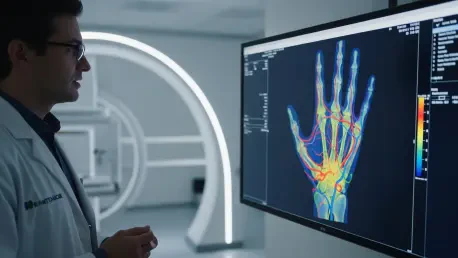
Recent proof-of-concept studies have demonstrated the impressive performance and broad applicability of the RUS-PAT system. Researchers have successfully imaged diverse regions of the human body, including the hand, foot, and breast, showcasing the technology’s versatility. The system’s imaging speed is a standout feature; it can capture a detailed 3D image of a region up to 10 centimeters wide in approximately 10 seconds. This efficiency contrasts sharply with the longer acquisition times required for many traditional imaging methods, such as MRI.
Furthermore, initial tests involving brain imaging have yielded promising results. These scans, conducted on patients undergoing surgery that required temporary skull removal, confirmed the system’s capacity to generate medically meaningful images of both brain tissue and its complex vascular networks. These benchmarks establish the technology’s potential to deliver high-quality, comprehensive 3D images with a speed and efficiency that could significantly improve clinical workflows and patient experiences.
Potential Clinical Applications Across Medical Fields
Advancements in Neurology and Brain Diagnostics
In the field of neurology, the hybrid system offers the potential for a new window into brain health. Its ability to visualize both tissue structure and blood flow could be instrumental in diagnosing and monitoring conditions such as stroke, where rapid assessment of blood supply is critical. Similarly, for traumatic brain injuries, the technology could provide immediate insights into the extent of tissue damage and associated vascular disruption, guiding urgent clinical interventions and improving patient outcomes.
Enhancing Oncology and Cancer Care
The applications in oncology are particularly compelling, especially for diagnosing and managing breast cancer. The ability to simultaneously visualize a tumor’s morphology and its associated blood supply provides a powerful diagnostic tool. The density and pattern of vasculature are often key indicators of a tumor’s aggressiveness and response to treatment. This dual-modality imaging could therefore enhance early detection, improve the accuracy of biopsies, and help clinicians tailor more effective treatment plans.
Transforming Vascular Medicine and Limb Preservation
This technology also holds immense promise for transforming vascular medicine. For the millions of individuals with diabetic foot complications, where poor circulation can lead to severe outcomes, RUS-PAT offers a rapid and low-cost method to image vasculature. By providing detailed maps of blood flow in the lower limbs, the system can help clinicians identify at-risk tissue, assess the severity of peripheral artery disease, and guide interventions aimed at preventing amputations and preserving limb function.
Current Limitations and Ongoing Research Efforts
The Challenge of Transcranial Imaging
Despite its potential, a primary technical hurdle for brain imaging is the intact human skull. Bone severely distorts and attenuates both light and sound signals, making it extremely difficult to obtain clear images of the brain noninvasively. This barrier currently limits brain scanning to situations where a portion of the skull is temporarily removed, such as during surgery.
To overcome this significant challenge, research efforts are focused on developing sophisticated solutions. Scientists are exploring the use of adjusted ultrasound frequencies that may penetrate bone more effectively. Concurrently, they are developing advanced computational algorithms designed to correct for the aberrations and signal loss caused by the skull, with the ultimate goal of enabling high-quality transcranial imaging.
Bridging the Gap from Prototype to Clinical Practice
As the technology is still in a proof-of-concept phase, further refinement is necessary to prepare it for widespread clinical deployment. A key focus of ongoing work is to improve the consistency and quality of the images produced across different scans and among diverse patient populations. Establishing robust and repeatable performance is a crucial step in validating the system as a reliable diagnostic tool. This refinement process will be essential for moving the technology from a promising prototype to a standard instrument in clinical practice.
Future Outlook and Long-Term Impact
The trajectory for hybrid photoacoustic ultrasound technology is pointed toward greater sophistication and broader accessibility. Future developments are expected to focus on enhancing its computational power, with advanced algorithms playing a key role in improving image reconstruction, correcting for artifacts, and expanding the system’s diagnostic capabilities. Further adjustments to ultrasound frequencies and laser parameters could also unlock new applications and improve imaging depth and resolution.
In the long term, this technology has the potential to become a standard, radiation-free diagnostic tool in mainstream clinical settings. Its combination of speed, safety, and comprehensive imaging could position it as a first-line diagnostic option in emergency rooms, outpatient clinics, and even at the patient’s bedside. As it matures, the RUS-PAT system may not only supplement existing imaging standards but could also replace them in scenarios where its unique advantages offer a superior clinical solution.
Concluding Assessment
The development of the hybrid photoacoustic ultrasound system marks a pivotal moment in medical imaging. The technology’s core strengths—its rapid acquisition speed, freedom from ionizing radiation, and unique ability to co-register anatomical structure with vascular function—collectively address many of the fundamental limitations of current diagnostic standards. This integrated approach provides a more complete and insightful picture of human biology in a single scan than was previously possible with conventional methods.
Overall, the RUS-PAT system represents a powerful and versatile platform with the potential to revolutionize diagnostics across numerous medical specialties. While further research and refinement are required to overcome existing hurdles and transition the technology from the laboratory to the clinic, its foundational principles have been firmly established. The initial performance benchmarks suggest that this hybrid imaging modality is on a path to becoming an indispensable tool for clinicians, promising to make advanced medical imaging safer, faster, and more accessible for patients worldwide.