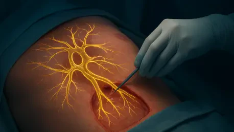

A collaborative team from MIT and Harvard has developed a technique called xvr to bridge the dangerous gap between real-time X-rays and preoperative 3D scans. Surgeons navigating the complex pathways of the human vascular system or delicate neurological structures often rely on two-dimensional fluoroscopy, which provides a flattened perspective of a multidimensional reality. This discrepancy creates a significant cognitive load, as clinicians must mentally reconstruct three-dimensional structures from grainy, shadows-only projections. The introduction of xvr represents a transformative leap in surgical navigation, utilizing artificial intelligence to synchronize these disparate data streams with unprecedented fidelity. By integrating high-resolution preoperative data directly into the live surgical feed, the system allows for a level of transparency that was previously unattainable in standard clinical environments. This advancement effectively addresses the critical need for spatial clarity during life-saving interventions such as acute stroke treatments or cardiac valve repairs.
Revolutionizing Image Alignment through Patient-Specific Modeling
Bridging the Dimensionality Gap: Part 1
The shift toward personalized medicine finds a powerful application in the xvr framework, which departs from the traditional reliance on generalized anatomical models that often fail to account for individual patient variance. Instead of training a single algorithm to recognize every possible human variation, this system generates a unique neural network for every procedure based on the specific patient’s own preoperative CT or MRI data. This process utilizes advanced physics-based simulations to create thousands of synthetic X-rays from virtually every conceivable angle within seconds. By calculating how X-ray photons interact with the specific density of that patient’s bone, soft tissue, and vascular structures, the AI gains a comprehensive understanding of the individual’s unique internal landscape. This high-fidelity simulation phase ensures that the resulting navigation tool is not just a generic map, but a precise, digital twin of the patient currently lying on the operating table.
Bridging the Dimensionality Gap: Part 2
One of the primary advantages of utilizing physics-based simulations rather than purely generative adversarial networks is the elimination of “AI hallucinations.” In many medical AI applications, there is a persistent risk that the algorithm might invent anatomical structures or hide pathologies based on patterns it learned from unrelated datasets. The xvr system avoids this pitfall by grounding its entire learning process in the actual physical properties of the patient’s scan. Since every synthetic image used for training is derived from a verified medical volume, the AI remains strictly tethered to anatomical reality. This level of reliability is paramount in high-stakes environments where even a millimeter of deviation can lead to vessel perforation or other surgical complications. Furthermore, the high speed of these simulations—generating upwards of 1,000 images per second—allows the system to be finalized just before the patient enters the operating suite, ensuring the most current data is always utilized for guidance.
Accelerating Preparation: Part 1
Despite the benefits of patient-specific modeling, the time required to train a deep-learning model from scratch has historically been a prohibitive barrier in emergency medicine. A standard training cycle for such complex neural networks can often take up to twelve hours, which is far too long for patients experiencing a time-critical event like an ischemic stroke. To circumvent this limitation, the researchers implemented a “foundation model” strategy. This involves a massive pre-training phase where the AI is exposed to a diverse library of whole-body 3D scans from over 2,000 individuals across various demographics and medical histories. This foundational knowledge provides the algorithm with a deep understanding of general human morphology and the physical laws governing X-ray attenuation. By the time a new patient arrives at the hospital, the AI already possesses a sophisticated baseline, allowing it to bypass the slow initial stages of the machine learning process.
Accelerating Preparation: Part 2
When a surgical team initiates the xvr protocol for a specific case, the system performs what is known as “fine-tuning,” adapting the existing foundation model to the patient’s specific scan in approximately five minutes. This rapid adaptation period transforms a generalized anatomical understanding into a bespoke navigation tool tailored to the exact curvature of a specific patient’s carotid artery or spinal column. Once this brief training phase is complete, the AI can perform real-time 2D-to-3D registration in a fraction of a second during the actual surgery. This speed is critical because it allows the system to update the surgical field visualization continuously, even if the C-arm X-ray machine is moved or the patient’s position shifts slightly. The ability to achieve sub-millimeter precision within such a condensed timeframe represents a significant engineering achievement that brings the power of advanced machine learning into the urgent atmosphere of the modern emergency department and trauma center.
Validating Clinical Efficacy and Expanding Medical Access
Empirical Success and Diverse Cases: Part 1
Validation of the xvr system involved an extensive comparison against both traditional manual methods and existing automated registration techniques. Researchers utilized the largest curated dataset of real-world 2D and 3D registrations, spanning five different medical institutions and covering a vast array of clinical scenarios. The results were consistent across the board, showing that the xvr system provided accuracy that was an order of magnitude superior to previous iterations of surgical AI. In neurovascular cases, where the margin for error is exceptionally thin, the system maintained sub-millimeter registration, ensuring that catheters could be guided through the brain’s delicate vasculature with absolute confidence. This high level of precision held steady across different body parts, including the pelvis, spine, and chest, proving that the technology is not limited to a single specialty but is instead a versatile platform for the entire field of interventional radiology.
Empirical Success and Diverse Cases: Part 2
The robustness of the algorithm was further demonstrated by its consistent performance across diverse patient populations, including pediatric and geriatric cases. Often, AI models trained on adult data struggle to adapt to the smaller scale and different bone densities found in children, yet the xvr foundation model approach effectively bridged this demographic gap. By learning from a wide spectrum of anatomical data, the system successfully navigated the unique challenges presented by developmental variations and age-related degenerative changes. This versatility ensures that the benefits of high-precision surgery are available to the most vulnerable patients who might otherwise face higher risks during invasive procedures. The ability to maintain such high standards of accuracy without requiring manual intervention from the surgical staff also reduces the cognitive burden on the team, allowing them to focus entirely on the nuances of the operative task.
Democratizing Care and Access: Part 1
The implementation of xvr has the potential to reshape the global healthcare landscape by mitigating the “geographical gap” that often limits access to specialized surgical care. Currently, many advanced minimally invasive procedures are only performed at large academic medical centers or specialized urban hubs because they require an extraordinary level of expertise in image interpretation and spatial navigation. For patients in rural or underserved areas, the time required for a transfer to one of these centers can lead to significantly worse outcomes, particularly in the case of stroke or heart attack where every second counts. By automating the most difficult aspects of surgical alignment and providing clear, 3D-guided visuals, xvr lowers the technical barrier for performing these complex interventions. This could eventually allow regional hospitals to offer the same level of precision and safety as their metropolitan counterparts, democratizing life-saving medical technology.
Democratizing Care and Access: Part 2
The development of the xvr technique demonstrated a successful synthesis of advanced machine learning and clinical necessity, proving that high-speed, patient-specific AI could realistically be integrated into the highest-stakes medical environments. By overcoming the dimensionality gap and the time constraints of traditional AI training, the research team established a new standard for surgical navigation that prioritized both speed and anatomical truth. Moving forward, the clinical community should focus on the widespread adoption of these foundation-model approaches to ensure that every operating room has access to the most sophisticated visualization tools available. Future efforts will likely concentrate on integrating these algorithms directly into medical imaging hardware, creating a seamless experience where the AI operates invisibly in the background. As the technology matured through 2026, it paved the way for a healthcare system that was more precise, more equitable, and fundamentally safer.