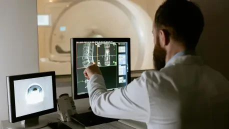
As a pioneer in the intersection of artificial intelligence and biomedical imaging, Laurent Giraid has dedicated his career to harnessing machine learning and innovative technologies to revolutionize clinical research. Today, we dive into his insights on MultiverSeg, a groundbreaking AI system designed to accelerate medical image segmentation. Our conversation explores how this tool streamlines a critical yet time-intensive process, its unique features that set it apart from existing solutions, and its potential to transform both research and healthcare practices with unprecedented efficiency.
Can you give us an overview of what MultiverSeg brings to the table for clinical researchers?
Absolutely. MultiverSeg is an AI-driven system developed to make the process of segmenting medical images much faster and more intuitive for researchers. Segmentation, which involves outlining specific regions of interest in images like brain scans or X-rays, is often a crucial first step in clinical studies. Our tool allows users to interact with the system through simple actions like clicking or scribbling on images, and it predicts the segmentation based on those inputs. What’s exciting is that as more images are processed, the system learns from them, eventually requiring no user input at all for new images. It’s a game-changer for speeding up research workflows.
Why is medical image segmentation such a critical yet challenging task in clinical research?
Segmentation is vital because it helps researchers quantify and analyze specific structures or abnormalities in medical images, like tracking changes in the brain’s hippocampus over time. However, it’s incredibly challenging because many structures are complex and hard to delineate accurately. Traditionally, this has been a manual process, which can take hours per image. For researchers juggling multiple tasks, segmenting just a handful of images a day can become a bottleneck, slowing down entire studies and delaying insights that could lead to medical breakthroughs.
How does MultiverSeg tackle the issue of time in the segmentation process?
MultiverSeg significantly cuts down on time by leveraging user interactions in a smart way. Initially, a researcher might click or scribble on an image to mark areas of interest, and the system uses those cues to predict the segmentation. As more images are segmented, the tool builds a context set—a kind of memory of previous work—and draws on that to improve its predictions. Over time, often by the ninth image or so, it might need just a couple of clicks, or none at all, to deliver accurate results. This incremental learning slashes the manual effort required.
Can you explain the role of the context set feature in enhancing MultiverSeg’s performance?
The context set is a core innovation in MultiverSeg. It stores previously segmented images and uses them as a reference to inform predictions on new images. Think of it as the system learning from experience—each segmented image adds to its knowledge base, allowing it to make more accurate guesses with less input from the user. The beauty of our design is that the context set can be any size; there’s no fixed number of images required for it to start working effectively. This flexibility makes it adaptable to a wide range of tasks and datasets.
What makes MultiverSeg stand out compared to other segmentation tools available to researchers?
Unlike many existing tools, MultiverSeg doesn’t require users to start from scratch with every new image or task. Traditional interactive tools demand repeated user input for each image, while task-specific AI models need hundreds of manually segmented images to train from the ground up. MultiverSeg bypasses both issues by using its context set to build on prior work and by not needing presegmented datasets or retraining. This means researchers can dive into new projects without extensive setup or machine-learning know-how, saving both time and resources.
Who is the ideal user for MultiverSeg, and how accessible is it for someone new to AI tools?
We designed MultiverSeg to be user-friendly for a broad audience, from clinical researchers to physicians. You don’t need to be an expert in machine learning to use it. The interface is intuitive—just upload an image and start marking areas of interest. There’s no need for extensive computational resources or complex setup. Whether you’re studying a new treatment or planning a clinical procedure, you can adapt the tool to your specific task with minimal effort, making it accessible even to those with limited technical background.
In practical terms, how do you see MultiverSeg impacting clinical research and healthcare delivery?
The potential impact is enormous. In research, MultiverSeg can accelerate studies on new treatments by reducing the time spent on segmentation, which could lower the costs of clinical trials and bring innovations to patients faster. In healthcare, it can support physicians in everyday tasks like planning radiation therapy, where precise segmentation of tumors or organs is critical. By automating and refining this process, the tool not only boosts efficiency but also enhances accuracy, ultimately improving patient outcomes and optimizing resource use in medical settings.
Looking ahead, what is your forecast for the role of AI tools like MultiverSeg in the future of biomedical imaging?
I believe tools like MultiverSeg are just the beginning of a transformative wave in biomedical imaging. As AI continues to evolve, we’ll see even greater integration into clinical workflows, with systems becoming more autonomous and capable of handling complex, multi-dimensional data like 3D images. The focus will likely shift toward real-time applications, where AI assists during live procedures or diagnostics. Additionally, ethical considerations and user feedback will shape these tools to ensure they’re trustworthy and tailored to real-world needs. The future is bright, with AI poised to bridge gaps between research and patient care like never before.


