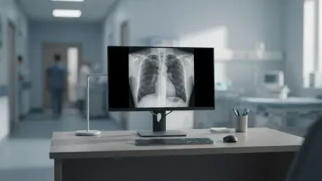

For identifying clinically significant effusions of 300 milliliters or greater, the AI achieved an Area Under the Curve of 0.85, demonstrating statistical non-inferiority to human specialists. Pleural effusion, the pathological accumulation of fluid within the pleural cavity, remains a significant clinical challenge across diverse medical scenarios, including congestive heart failure, pneumonia, and malignancy. The accuracy of fluid volume assessment is paramount in determining whether a patient can be managed through conservative monitoring or if they require immediate invasive procedures like thoracentesis or chest tube placement. While chest radiographs serve as the primary diagnostic tool due to their accessibility and low radiation dose, their two-dimensional nature presents a complex geometric puzzle when clinicians try to infer three-dimensional fluid volumes. This limitation has historically led to variability in human interpretation, prompting researchers to explore the potential of multimodal artificial intelligence models to standardize and enhance the quantification of pleural fluid.
Evaluating Diagnostic Accuracy and Methodology
Defining the Gold Standard: CT Volumetry
The research was designed as a retrospective observational study at Motol and Homolka University Hospital, focusing on 88 adult patients who underwent both chest radiographs and thoracic CT scans within a very tight window. This three-hour median interval was critical to ensure that physiological fluid shifts or medical interventions did not alter the volume between the two imaging sessions. The study analyzed a total of 176 hemithoraces, providing a robust dataset for comparing traditional interpretative methods with modern computational analysis. By utilizing CT scans with slice thicknesses of five millimeters or less, the team ensured that the data used as the “gold standard” was as precise as current medical technology allows. This rigorous selection process provided a solid foundation for evaluating the accuracy of both human radiologists and the multimodal AI model in a real-world clinical environment.
To establish the reference standard, specialized software was used to perform manual segmentation of the fluid on axial CT reconstructions. A radiology resident meticulously contoured the fluid levels, which were then verified through inter-slice extrapolation to calculate the total volume in milliliters. This manual process was not only time-intensive but was also subjected to rigorous validation, including independent verification by a board-certified radiologist after a four-week washout period. The resulting intraclass correlation coefficient of 0.98 for both intra-observer and inter-observer reliability confirmed that these volumetric measurements were an exceptionally stable ground truth. Such a high degree of precision in the reference standard allowed the researchers to definitively attribute any discrepancies in the radiograph assessments to the limitations of the projection imaging itself or the diagnostic process of the observer, rather than to errors in the baseline measurement.
Implementing the AI: Human Assessment Comparison
The human element of the study involved two board-certified thoracic radiologists with 9 and 19 years of professional experience, respectively. These specialists were tasked with independently reviewing the radiographs and providing volumetric estimates based on established radiographic markers, such as the meniscus sign and diaphragm obscuration. In addition to their volume estimates, the radiologists recorded their confidence levels for each hemithorax using a five-point Likert scale. To ensure their assessments were reliable, their repeatability was tested, revealing high internal consistency. This established a human performance ceiling, reflecting the best possible outcomes currently achievable by specialists using standard 2D imaging. The involvement of experienced clinicians ensured that the AI would be compared against a high level of diagnostic expertise rather than a generalized or less-experienced baseline.
In early 2026, several leading multimodal models were prescreened for this quantitative task, with ChatGPT 5.2 ultimately selected as the most capable based on preliminary performance metrics. The AI was provided with high-resolution screen captures of the radiographs and guided by a “zero-shot” prompt that instructed it to adopt the persona of a thoracic imaging expert. Unlike traditional AI models that require thousands of labeled training images, this model utilized its broad training to reason through the visual data and provide volumetric estimates for both sides of the chest. The AI repeated its analysis three times within a single session to demonstrate internal consistency, achieving a nearly perfect correlation coefficient. This phase of the study highlighted the transition toward general-purpose multimodal models that can perform specialized medical tasks without the need for bespoke software development or extensive fine-tuning.
Comparative Performance and Clinical Implications
Statistical Parity: AI and Human Experts
The core finding of the study revealed that all three evaluators—the two radiologists and the AI—achieved a moderate level of agreement with the CT-derived reference standard. The statistical analysis showed that the AI’s intraclass correlation coefficient of 0.66 was remarkably close to the humans’ scores of 0.64 and 0.74. This parity suggests that modern multimodal AI models have developed the capacity to process visual medical data and reach conclusions that are statistically indistinguishable from those of highly trained human specialists. While “moderate” agreement might seem lower than desired, it reflects the inherent difficulty of the task; no observer, human or machine, can perfectly reconstruct 3D volumes from a 2D shadow. The fact that the AI hit the same performance level as the human experts indicates it has reached the physical limits of what is possible with radiographic technology.
Beyond simple correlations, the performance similarity between the AI and human experts has profound implications for future clinical workflows. If a general-purpose model can provide estimates that align with those of board-certified thoracic radiologists, it could serve as a valuable diagnostic support tool in settings where specialists are not immediately available, such as emergency departments or rural clinics. This parity does not suggest that the AI is better than the human, but rather that it is equally capable of navigating the geometric complexities and ambiguities of projection imaging. As healthcare systems move toward more integrated digital environments, having a tool that provides a reliable “second opinion” or an automated preliminary assessment could significantly reduce the cognitive load on radiologists and help prioritize cases that require more urgent cross-sectional imaging for definitive management.
Identifying Risk: The 300 Milliliter Threshold
One of the most clinically relevant aspects of the study was the evaluators’ ability to identify effusions exceeding 300 milliliters, which is often the threshold for considering therapeutic drainage. The AI’s performance in this specific area was particularly impressive, achieving an Area Under the Curve of 0.85. This metric indicates that the model is highly effective at discriminating between small, clinically insignificant fluid collections and larger ones that likely require intervention. While one radiologist performed slightly better with a score of 0.90, the AI was statistically non-inferior to the other specialist. This suggests that for the practical purpose of triage, the AI is a reliable tool for identifying patients who are at higher risk and may need immediate attention or further diagnostic testing to confirm the exact fluid volume before a procedure.
The ability to accurately flag significant effusions has direct consequences for patient safety and resource allocation. In a busy hospital setting, quickly identifying which patients require specialized procedures like thoracentesis can prevent delays in treatment and improve overall outcomes. The AI’s performance at the 300-milliliter mark shows that it can handle the nuances of fluid distribution and density on a radiograph well enough to serve as a screening mechanism. By automating the identification of these significant cases, the AI can help clinicians focus their attention on the most critical patients. This shift toward quantitative screening, rather than just binary presence-or-absence detection, represents a major step forward in the evolution of medical AI, providing clinicians with actionable data that mirrors the decision-making process used in daily practice.
Factors Affecting Interpretive Success
Positional Impact: Supine versus Upright Results
The research identified that the orientation of the patient during the X-ray significantly impacted the accuracy of the volumetric estimations for both the AI and the human experts. In the study cohort, radiographs were taken in various positions, including standing, sitting, and supine, mirroring the reality of clinical practice where critically ill patients often cannot remain upright. The AI encountered more significant challenges with supine radiographs, where pleural fluid tends to layer posteriorly along the chest wall. This layering creates a diffuse “haze” or a generalized increase in opacity rather than the clear, curved meniscus sign typically seen in upright images. Consequently, the lack of distinct anatomical markers in the supine position makes it much more difficult to infer the true volume of the fluid from a two-dimensional projection.
This positional difficulty highlights a fundamental limitation of thoracic imaging that affects any interpreter, regardless of their nature. The loss of three-dimensional data during the projection process creates a “ceiling” of potential accuracy that cannot be easily bypassed without switching to a different imaging modality like ultrasound or CT. For the AI, the increased error in supine positions suggests that while it is highly capable of geometric reasoning, it still struggles with the same visual ambiguities that plague human observers. Recognizing these limitations is crucial for clinicians who might rely on AI-generated reports; an estimate from an upright X-ray is likely to be more accurate than one derived from a supine bedside film. Understanding where the AI is most likely to falter allows for better integration of these tools into the broader diagnostic strategy, ensuring they are used where they are most effective.
Confidence Scores: The Accuracy Correlation Paradox
A particularly intriguing discovery in the research was the total lack of correlation between an interpreter’s confidence and their actual accuracy. Both the radiologists and the AI were asked to provide a confidence score for their estimates, yet a high level of certainty did not lead to a more precise volume calculation. This phenomenon, often referred to as the overconfidence effect, suggests that the subjective feeling of diagnostic certainty is often detached from the objective truth of the case. In many instances, an evaluator might feel very confident because they see a clear meniscus, yet the actual volume might be significantly different due to hidden fluid pockets or unusual chest wall anatomy. This paradox highlights a significant risk in clinical practice, where a high-confidence but incorrect report could lead to inappropriate medical decisions.
For the AI, this lack of correlation indicates that even though the model can provide a consistent and seemingly authoritative answer, it does not yet have a calibrated “meta-awareness” of its own limitations in complex visual tasks. This insight is vital for the development of future medical AI systems, as it emphasizes the need for objective performance metrics rather than subjective probability scores. Clinicians must remain skeptical and use AI estimates as one piece of a larger diagnostic puzzle rather than as a definitive measurement. The finding encourages a more cautious approach to interpretation, where the focus is shifted from how certain an observer feels to the evidence-based limitations of the imaging modality itself. This understanding reinforces the role of AI as a collaborative tool that requires human oversight to navigate the nuances of patient-specific anatomical variations.
Future Directions in Multimodal AI
General Intelligence: Zero-Shot Reasoning Capabilities
The success of ChatGPT 5.2 in this study marks a pivotal shift in medical technology, as it demonstrated sophisticated quantitative reasoning without being specifically trained on thousands of labeled pleural effusion images. Traditional AI models used in radiology are typically “narrow” systems, designed and trained for a single task, such as detecting a lung nodule or identifying a fracture. In contrast, the multimodal AI used here relied on “zero-shot” learning, applying its general understanding of geometry, anatomy, and physics to a specialized task it had not been specifically optimized for. This ability to generalize across domains suggests that we are moving toward a future where a single, versatile AI model could support a wide range of radiological tasks, significantly reducing the cost and complexity of deploying AI in clinical settings.
The use of general-purpose models also allows for a more natural interaction between the radiologist and the software through the use of natural language prompts. Instead of interacting with a rigid user interface, clinicians can provide context or ask specific questions about the images, much like they would when consulting with a colleague. This flexibility could lead to much faster innovation, as researchers can test new diagnostic queries without needing to rebuild or retrain the underlying model. While specialized models will likely remain superior for certain high-precision tasks, the performance of multimodal AI in this study suggests that general intelligence is becoming a viable and powerful tool for complex medical inference. As these models continue to evolve, their ability to integrate visual data with textual clinical information will likely become a cornerstone of the next generation of diagnostic support systems.
Clinical Integration: The Evolving Radiological Workflow
The findings from this study confirm that while AI has reached parity with human specialists in volume estimation, it is not a replacement for the high-precision measurements provided by CT or ultrasound. Instead, the current value of AI lies in its role as a collaborative partner that can provide rapid, automated “second opinions” in the early stages of the diagnostic process. By integrating these models into the radiological workflow, healthcare systems can ensure that every radiograph receives a baseline quantitative assessment, which can help in prioritizing worklists and identifying critical findings that might otherwise be overlooked during a busy shift. This collaborative approach leverages the consistency and speed of AI while maintaining the essential oversight and nuanced judgment provided by experienced human radiologists.
In the final analysis, the integration of multimodal AI into clinical practice should be viewed as a forward-looking strategy to overcome the physical limitations of traditional 2D imaging. Future developments in “prompt engineering” could further refine these models by providing them with additional patient-specific context, such as body mass index, medical history, or the specific clinical question being asked. This holistic approach could potentially narrow the error margins that currently affect both humans and machines. The transition from simple detection to complex quantification is now well underway, and as these tools become more calibrated and reliable, they will play an increasingly central role in clinical decision-making. The goal is to move toward a more integrated, data-driven diagnostic environment where AI supports clinicians in providing the most accurate and timely care possible for patients with complex conditions.