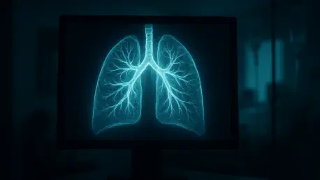

Microscopic lung vulnerabilities that escape human detection during routine staging are now being mapped by self-supervised learning algorithms to enhance patient safety protocols. The advent of immune checkpoint inhibitors has fundamentally altered the landscape of cancer treatment by enabling the immune system to recognize and destroy tumor cells. While these therapies have provided long-term survival for many, they often come with a high physiological cost known as checkpoint-inhibitor pneumonitis. This inflammatory lung condition affects roughly 10% of patients and can quickly escalate into a life-threatening emergency if not caught early. In a major step forward, researchers at The University of Texas MD Anderson Cancer Center developed the CIPHER model, which uses artificial intelligence to scan routine, pretreatment CT images for subtle warning signs. Identifying these sub-visual signatures allows clinicians to predict which individuals are most likely to experience complications.
Development of the CIPHER Foundation Model
The CIPHER tool represents a significant departure from previous radiomics approaches that relied on narrow, pre-programmed markers of disease. Instead, this system was built as a foundation model using self-supervised learning, a technique that allows the AI to learn from vast amounts of data without explicit human labeling. During its initial development phase, the algorithm processed over 590,000 individual CT image slices from a diverse database of 2,500 lung cancer patients. This intensive training period enabled the model to internalize the complex structural hierarchy of the human lung, mapping the intricate networks of blood vessels, airways, and various textures of healthy parenchyma. By mastering the nuances of normal and abnormal anatomy first, the AI established a deep understanding of pulmonary health that far exceeds the analytical capacity of traditional software. This method ensured the model was grounded in biological reality rather than simple patterns.
Once the foundational understanding of lung anatomy was established, the research team, led by experts in radiation oncology and thoracic medicine, fine-tuned the model to differentiate between patients who eventually developed pneumonitis and those who did not. This two-step process of learning the basic language of medical imaging before applying it to a specific clinical prediction is what allows the system to identify future risks that are invisible to the human eye. By analyzing the subtle textures and density variations in pretreatment scans, the AI can detect pre-existing microscopic inflammation or tissue damage that suggests a predisposition to adverse reactions. This transition from general anatomical knowledge to predictive clinical insight represents a major leap in how machine learning is applied to oncology. It moves the technology beyond simple diagnostic classification and into the realm of truly personalized risk assessment, providing a crucial safety net for high-risk patients.
Predictive Performance and Statistical Accuracy
To confirm the reliability of these findings, the effectiveness of the AI was tested using pretreatment scans from a cohort of patients with non-small cell lung cancer. The researchers validated the model against an independent external dataset to ensure the results were not limited to a single institution or specific patient population. In both the internal and external groups, the model achieved a high statistical accuracy with an Area Under the Curve of approximately 0.83. This level of performance indicates a robust and highly reliable predictive capability, as an AUC of 1.0 represents a perfect prediction. Crucially, the system significantly outperformed traditional risk-assessment methods that rely on clinical factors like age or smoking history. It also proved more accurate than conventional radiomics, which uses hand-designed quantitative features. This success highlights the power of self-supervised learning in capturing complex biological signals that human experts miss.
A common hurdle for medical AI is the tendency to perform well on training data but fail in real-world settings due to differences in scanner types and imaging protocols. However, this model demonstrated remarkable resilience when faced with varying technical conditions. Even when the external validation group utilized different CT hardware and imaging techniques, the algorithm maintained its predictive accuracy without significant degradation. This stability proves that the AI is identifying genuine biological indicators of lung vulnerability rather than reacting to technical artifacts or population-specific noise. Such resilience is essential for any tool intended for widespread clinical adoption across different healthcare systems with diverse equipment. By overcoming the problem of overfitting, the researchers have created a system that can be reliably deployed in various hospital environments. This robustness ensures that the predictions remain consistent regardless of where a patient receives their initial staging scans.
Clinical Implications for Personalized Oncology
The ability to identify high-risk patients before the first dose of immunotherapy is administered offers a transformative opportunity for personalized oncology. The study revealed that patients flagged by the AI as high-risk tended to develop pneumonitis earlier in their treatment course than others. This finding suggests that the model measures a spectrum of lung fragility rather than providing a simple binary outcome. By understanding where a patient falls on this spectrum, oncologists can make more informed decisions about the frequency of clinical follow-ups. For those in the highest risk categories, doctors might implement more intensive monitoring schedules or use proactive imaging to catch early signs of inflammation before they become symptomatic. This shift toward personalized risk stratification allows for a more efficient use of medical resources while significantly reducing the likelihood of a patient suffering a catastrophic respiratory event during their cancer treatment.
Furthermore, the model’s predictions remained statistically significant even after the researchers adjusted for known clinical variables that contribute to lung injury, such as prior radiation therapy to the chest. Radiation is a well-known risk factor for lung inflammation, yet the AI provided additional predictive value beyond what radiation history alone could offer. This indicates that the tool is detecting microscopic tissue vulnerabilities that currently escape traditional clinical detection methods. From a practical standpoint, the implementation of such a tool allows oncologists to prioritize high-risk patients for enrollment in clinical trials aimed at preventing immunotherapy-related toxicities. In cases where the predicted risk is exceptionally high, clinicians might even reconsider the intensity of the immunotherapy or combine it with protective measures. This proactive approach ensures that the pursuit of tumor eradication does not come at the expense of long-term pulmonary health.
Future Directions and Broad Applications
While the current success of the model is centered on lung cancer, the research team is actively working to expand its scope to other malignancies. Because immune checkpoint inhibitors are now the standard of care for various conditions, including melanoma, kidney cancer, and bladder cancer, there is a pressing need for universal predictive tools. Future studies will focus on integrating the model into real-time clinical workflows, allowing it to run automatically in the background as staging scans are processed. This would ensure that risk scores are available to oncologists immediately, without requiring additional manual steps or specialized imaging procedures. By 2027 and beyond, the goal is to refine these algorithms so they can function across a wide range of cancer types and treatment combinations. Scaling this technology across the oncology spectrum represents the next major milestone in the quest to minimize the side effects of powerful new immune-stimulating drugs.
The development of the CIPHER system demonstrated that routine medical imaging held a wealth of untapped data capable of transforming patient safety standards. Clinicians who integrated these AI-driven insights into their practice successfully transitioned from a reactive treatment model to a proactive prevention strategy. The research suggested that future implementation efforts should focus on creating automated background processes that analyze staging scans without adding to the radiologist’s manual workload. Furthermore, the expansion of the model to include multi-modal data, such as blood markers and genetic signatures, provided a comprehensive roadmap for managing the complexities of modern immunotherapy. Hospitals that prioritized the deployment of these predictive tools saw a measurable improvement in the management of adverse events, ultimately making life-saving treatments safer for a broader population of patients. This shift in oncology practice established a new benchmark for precision medicine.