
Diagnostic imaging is currently undergoing a radical transformation as clinical practitioners struggle to keep pace with a global surge in chronic disease and a shrinking workforce. The traditional ultrasound exam, long dependent on the manual dexterity and subjective interpretation of a sonographer, is being reimagined through the lens of artificial intelligence. GE HealthCare’s latest LOGIQ portfolio—featuring the E10 Series, Fortis, and Totus—arrives at this critical juncture, promising to turn raw acoustic data into actionable clinical intelligence. This shift is not merely about sharper images; it represents a fundamental change in how medical professionals interact with hardware to make life-saving decisions under pressure.
Evolution of Intelligent Ultrasound Imaging
The journey toward intelligent imaging began with the necessity to standardize quality across varying levels of user expertise. In the past, the disparity between a novice and an expert sonographer could lead to inconsistent diagnostic outcomes, particularly in complex abdominal cases. Modern systems have evolved to address this by moving beyond simple pixel processing to integrated algorithmic support. The current generation of technology leverages deep learning to recognize anatomical structures in real-time, effectively providing a digital “second set of eyes” that monitors the exam as it happens.
This evolution is situated within a broader technological landscape where data liquidity and interoperability are paramount. Ultrasound is no longer a siloed diagnostic tool but a connected node within a hospital’s digital architecture. By shifting from closed proprietary systems to open platforms like Verisound, manufacturers are allowing for a more modular approach to medical software. This context is essential because it means the value of the hardware is now inextricably linked to its ability to host and execute complex AI models that can be updated as medical knowledge expands.
Core Technical Features and Smart Components
AI-Driven Automation and Workflow Optimization
One of the most impactful features of the new LOGIQ series is the Auto Abdominal Suite 2.0. This component functions by utilizing sophisticated neural networks to identify and segment organs automatically. For instance, the Auto Aorta and Auto CBD (Common Bile Duct) assistants can reduce manual system interactions by up to 80%. This is significant because it mitigates the repetitive stress injuries common among sonographers while simultaneously accelerating the exam process by roughly 65%. Instead of clicking and dragging calipers, the clinician simply confirms the measurements the AI suggests.
Moreover, the performance of these automated tools remains consistent even when dealing with difficult patient habitus. The system’s ability to maintain high-resolution segmentation in technically challenging cases is what differentiates it from basic automation found in lower-tier models. This level of optimization ensures that the “time-to-insight” is minimized, allowing high-volume clinics to maintain throughput without sacrificing the depth of the clinical evaluation. It marks a transition from manual labor to supervisory interpretation.
Advanced Transducer Integration and Hardware Synergy
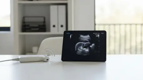
The hardware component of these systems has undergone a parallel revolution, specifically regarding the synergy between traditional console probes and wireless technology. The integration of the Vscan Air into the high-end LOGIQ ecosystem allows for a hybrid workflow that was previously impossible. A clinician can now switch from a deep-tissue console probe to a handheld wireless transducer for quick vascular checks or superficial scans without interrupting the patient session. This flexibility ensures that the imaging environment adapts to the patient’s immediate needs rather than forcing the patient to conform to the machine’s limitations.
Technical performance is further bolstered by the use of wide-band transducers that provide exceptional detail from the near to the far field. This hardware synergy is critical for advanced applications like elastography or fat fraction measurement, where the quality of the raw signal determines the accuracy of the quantitative data. By pairing high-performance piezoelectric materials with digital beamforming, these systems capture more nuance in tissue texture, which the AI then uses to provide more precise diagnostic indicators.
Current Industry Trends and Digital Ecosystems
A defining trend in the current medical landscape is the move toward “platformization.” Modern ultrasound systems are increasingly built on open digital architectures that support third-party application integration. This shift reflects a change in consumer behavior among healthcare providers who now prioritize longevity and scalability over static hardware specifications. By enabling on-scanner reporting and cloud-based data sharing through tools like ViewPoint Ultra, these systems bridge the gap between the imaging suite and the physician’s office, ensuring that data is accessible wherever it is needed.
Furthermore, there is a growing emphasis on semi-quantitative imaging. The industry is moving away from purely visual assessments—which are prone to inter-operator variability—and toward data-driven metrics. The emergence of tools that measure liver fat burden or tissue stiffness is a direct response to the global rise in metabolic disorders. This trend underscores a broader shift in healthcare toward personalized medicine, where the ultrasound system serves as a sophisticated biophysical sensor capable of tracking disease progression over time with mathematical precision.
Real-World Clinical Applications and Sector Impact
The most profound impact of this technology is seen in the field of hepatology. With metabolic dysfunction-associated fatty liver disease affecting nearly 40% of the population, the introduction of the Ultrasound-Guided Fat Fraction (UGFF) tool has become a game-changer. This application provides a non-invasive, quantifiable method for monitoring liver health that is far more cost-effective than MRI. In large-scale clinical settings, this allows for the early identification of fibrosis or steatosis, potentially preventing the progression to more severe chronic conditions.
Beyond specialized liver care, these systems are making a significant mark in emergency and general diagnostic departments. In high-pressure environments, the ability of the AI to standardize measurements means that a trauma surgeon or a general practitioner can obtain reliable data quickly. This democratization of high-end diagnostic capability ensures that a consistent standard of care is maintained regardless of which department the patient enters. The unique implementation of AI assistants across different clinical “suites” makes the technology versatile enough for everything from vascular studies to complex urology.
Technical Barriers and Adoption Challenges
Despite these advancements, the path to widespread adoption is not without hurdles. One of the primary technical barriers is the “black box” nature of some AI algorithms, which can lead to skepticism among seasoned clinicians. There is a learning curve associated with trusting automated measurements, and regulatory bodies remain cautious about the autonomy granted to diagnostic software. Additionally, the integration of third-party apps on an open platform raises valid concerns regarding data security and patient privacy, necessitating robust cybersecurity frameworks that must be constantly updated.
Market obstacles also include the high initial capital expenditure required to upgrade to “intelligent” systems. While the long-term efficiency gains are clear, many smaller clinics struggle with the cost of hardware that requires frequent software subscriptions to remain “smart.” To mitigate these limitations, development efforts are currently focused on creating more modular upgrade paths, allowing facilities to add specific AI capabilities as their budget and clinical needs evolve, rather than requiring a total system overhaul.
Future Outlook and Technological Trajectory
Looking ahead, the trajectory of ultrasound technology points toward even greater levels of predictive analytics. We are moving toward a future where the system will not only measure what is there but will also use historical patient data to suggest potential diagnostic paths. The integration of generative AI could eventually allow for the creation of synthetic anatomical models based on a patient’s specific scans, helping surgeons plan interventions with a level of spatial awareness that was previously unattainable.
Furthermore, the decentralization of imaging will likely accelerate. As AI algorithms become more efficient, they will be deployed on increasingly smaller devices, potentially making the diagnostic power of a LOGIQ system available in a pocket-sized format. This will shift the role of the hospital-grade console toward becoming a centralized hub for complex data synthesis, while the actual point-of-care imaging happens at the bedside, in the ambulance, or even in the patient’s home.
Assessment of the Intelligent Imaging Landscape
The transition to AI-powered ultrasound represents a successful synthesis of hardware reliability and software intelligence. The current state of the technology proves that automation can effectively address the twin challenges of clinician burnout and rising patient volumes without compromising diagnostic integrity. By quantifying previously qualitative observations, these systems have moved ultrasound into the realm of precision medicine, providing a more objective basis for patient management and long-term monitoring across a variety of medical sectors.
Ultimately, the impact of these intelligent systems was measured by their ability to disappear into the background of clinical practice, allowing the physician to focus entirely on the patient. The strategic move toward open digital platforms ensured that these investments remained relevant even as new diagnostic challenges emerged. As healthcare systems continue to adapt, the successful implementation of these tools suggested that the future of medicine would be defined not by the quantity of data collected, but by the speed and accuracy with which that data could be turned into a cure.