
European medical technology company AZmed has officially announced the commercial launch of AZnod, a CE-marked artificial intelligence tool poised to significantly impact the landscape of lung cancer screening. This strategic move signals the company’s expansion from its established expertise in standard X-ray analysis into the more data-intensive domain of computed tomography (CT) imaging. As the inaugural product in AZmed’s newly unveiled Rayscan® product line, AZnod is a sophisticated software solution engineered to automate the detection and comprehensive characterization of pulmonary nodules. The technology is designed to provide critical support to radiologists, addressing the growing challenges posed by high-volume screening programs and aiming to enhance both the speed and accuracy of early cancer detection, a pivotal factor in improving patient outcomes. This launch represents a significant step forward in applying AI to one of the most pressing areas of modern diagnostic imaging.
Addressing the Rising Demands of Modern Screening
The global proliferation of lung cancer screening programs, while vital for early detection, has inadvertently created a significant operational and interpretive strain on medical imaging departments. Radiologists are now tasked with evaluating an unprecedented volume of low-dose CT examinations, a process that demands meticulous attention to detail. The core of this challenge lies in the identification of small, early-stage pulmonary nodules, which can manifest in various forms, including solid, part-solid, or pure ground-glass opacities. Subsolid nodules, in particular, present a considerable diagnostic hurdle as they can be difficult to visualize yet are clinically significant. The ability to detect these subtle indicators at the earliest possible stage is paramount, as it has been proven to substantially improve patient survival rates. AZnod is positioned as a direct response to this escalating workflow pressure, offering a technological solution to augment the capabilities of clinicians on the front lines of cancer detection.
Furthermore, a critical aspect of effective screening programs is the standardization and reproducibility of radiological reports. Clinicians are required to deliver assessments that are consistent and strictly aligned with established clinical frameworks, such as the Lung-RADS (Lung Imaging Reporting and Data System) and the Fleischner Society guidelines. Adherence to these standards is essential because even minor variations in the measurement of a nodule’s dimensions or volume can drastically alter a patient’s management plan, potentially leading to different follow-up schedules or treatment pathways. The subjective nature of manual measurement can introduce inter-observer variability, creating inconsistencies that can impact patient care. AZnod is engineered to mitigate these challenges by providing objective, automated analysis, thereby enhancing the efficiency of the screening process and promoting greater consistency in diagnostic output across different readers and institutions.
The Functional Core of the AZnod System
The AZnod AI tool is meticulously engineered to serve as a powerful assistant to the radiologist, automating key facets of nodule analysis without disrupting established diagnostic procedures. The system’s operation commences with a comprehensive analysis of every individual slice within a given CT scan, a granular approach that ensures no section of the lung parenchyma is overlooked. Its sophisticated detection algorithm is specifically calibrated to identify nodules that fall within the clinically relevant size range of 3 to 30 millimeters. Once a potential nodule is identified, AZnod executes a detailed evaluation, providing a suite of standardized, quantitative measurements. This includes the nodule’s volume, expressed in cubic millimeters, as well as its long-axis and perpendicular diameters. By generating these precise and reproducible metrics, the tool directly addresses and helps to mitigate inter-observer variability, a persistent challenge in manual radiological assessment that can affect diagnostic accuracy and consistency.
A key differentiator of AZnod lies in its advanced characterization capabilities, which extend far beyond simple detection and measurement. The AI model performs a sophisticated classification of each detected nodule based on a set of critical diagnostic attributes. This detailed characterization encompasses the nodule’s density, its contour morphology—distinguishing between smooth, irregular, or spiculated borders—its internal composition, such as solid, cystic, or calcified components, and its precise anatomical positioning within the lungs. This multi-faceted analysis provides the rich, structured data necessary to support a guideline-aligned clinical assessment. By automating this complex part of the diagnostic process, AZnod facilitates a more objective and streamlined reporting workflow, allowing radiologists to focus their expertise on interpretation and clinical decision-making, ultimately leading to a more robust and efficient diagnostic pathway for the patient.
Seamless Integration and Strategic Significance
A fundamental aspect of AZnod’s design is its seamless integration into existing clinical environments, a feature critical for widespread adoption. The system is built to process images that are strongly pseudonymized, directly addressing crucial patient data privacy and security concerns from the outset. Following its detailed analysis, the tool delivers a structured radiological output directly to the user’s existing platform. This output is thoughtfully consolidated into a single, comprehensive report designed for maximum clarity and efficiency. The report intelligently organizes all findings by listing each detected nodule according to its clinical priority, a feature that enables the radiologist to immediately focus their attention on the most suspicious or significant lesions. This prioritized presentation is a key element in optimizing the review process and ensuring that critical findings are not overlooked in complex cases with multiple nodules.
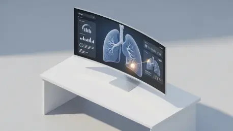
The report generated by AZnod provides a wealth of information in an easily digestible format. Each entry is accompanied by annotated views that visually highlight the nodule and prominently display its standardized measurements, offering immediate visual confirmation. This is complemented by a detailed list of the nodule’s diagnostic attributes as determined by the AI. A significant and user-friendly feature is the inclusion of an anatomical lung schematic, which provides a quick, intuitive visual map of where all detected nodules are located within the pulmonary system. This combination of prioritized lists, detailed metrics, and clear visual aids was intended to allow for a more rapid and confident assessment of nodules. The availability of AZnod for adoption by healthcare providers in all CE markets opened a major European market, reinforcing AZmed’s mission to leverage AI to improve diagnostic consistency and enhance operational efficiency within the more than 2,500 healthcare facilities it already served. This introduction was a calculated move to provide a tangible solution to the pressing challenges of high-volume lung cancer screening.