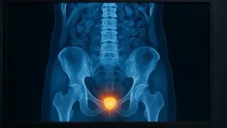

The search for a virtual biopsy alternative has led to the development of AI models that analyze gray-level co-occurrence and run-length matrices within standard CT images. This methodology is currently revolutionizing how oncologists approach bladder cancer, a disease that remains one of the most persistent threats within the urinary system. By leveraging machine learning, researchers have found a way to bypass the physical limitations of traditional tissue sampling, which often fails to capture the full genetic diversity of a tumor. A recent multi-institutional study has successfully demonstrated that routine computed tomography urography contains enough hidden data to accurately predict a patient’s HER2 status. This protein is a vital biomarker for determining eligibility for high-end therapies like antibody-drug conjugates. This discovery bridges the gap between conventional radiology and advanced molecular profiling, offering a clear path toward a more efficient diagnostic era where data takes center stage in treatment.
The Technological Foundation of Radiomics
Medical professionals have long relied on visual interpretation, but the advent of radiomics transforms pixels into quantitative metrics that define tumor biology. In this specific research, a cohort of 190 patients underwent a specialized CT urography protocol that captured the urinary tract across three distinct phases. These windows of observation allow clinicians to see how contrast moves through the tumor, revealing details about vascularity and tissue density that remain invisible to the naked eye. By extracting hundreds of features related to shape, intensity, and spatial complexity, the research team created a digital fingerprint of the malignancy. These mathematical descriptors go beyond simple diameter or volume, delving into the micro-scale distribution of gray levels within the image. This level of detail is essential for identifying the subtle heterogeneity that characterizes bladder cancer, providing a comprehensive view that a single needle biopsy often misses during a routine diagnostic workup.
Managing the immense volume of data generated by such high-throughput extraction is a significant challenge, as models can easily fall into the trap of overfitting. To combat this, the researchers implemented the Least Absolute Shrinkage and Selection Operator, commonly known as LASSO regression. This statistical technique acts as a filter, discarding redundant or noisy features and retaining only those with the highest predictive value for HER2 expression. By narrowing down the data to a select group of robust indicators, the scientists ensured that the resulting AI model would remain generalizable across different patient groups rather than just memorizing a specific dataset. This refinement process is critical for moving machine learning from an experimental phase into a tool that can be reliably deployed in a hospital setting. The reduction of variables also enhances the transparency of the model, allowing developers to understand which specific radiographic textures are most strongly associated with the underlying molecular profile.
Evaluating Machine Learning Classifiers
The performance of the predictive system was evaluated by splitting the patient population into two distinct groups, ensuring that the machine learning models were tested on data they had never encountered before. Five different classification algorithms—Logistic Regression, Support Vector Machine, K-Nearest Neighbors, XGBoost, and Random Forest—were put through their paces to see which could most accurately identify HER2-positive cases. The primary metric for success was the Area Under the Receiver Operating Characteristic Curve, where a score of 1.0 represents perfect diagnostic accuracy. This rigorous testing environment simulated real-world conditions, where a diagnostic tool must perform consistently across a diverse range of patient ages and tumor types. Each algorithm brought a different mathematical logic to the problem, from the simple linear approach of regression to the complex decision trees of the Random Forest, providing a broad spectrum of analysis to ensure the most reliable path to a non-invasive molecular diagnosis.
The results revealed a clear winner in the quest for imaging precision, as the Random Forest model outperformed its peers, especially when analyzing images from the corticomedullary phase of the CT scan. This specific timing, which captures the early arterial blood flow, yielded an AUC of 0.798 in the training set and 0.734 in the validation set. This finding is particularly significant because it underscores the importance of vascular timing in oncological imaging. High HER2 expression seems to alter the microvascular architecture of the bladder tumor in a way that is most visible during this brief window of peak enhancement. By focusing on this phase, the AI was able to detect the signature patterns of protein overexpression without the need for additional specialized scans. This efficiency proves that modern diagnostic equipment is already capable of providing high-level molecular insights if the data is analyzed through the right algorithmic lens, essentially turning a standard procedure into a screening tool.
Enhancing Accuracy Through Combined Data
A pivotal realization in this research was that artificial intelligence reaches its maximum potential when it works alongside established medical knowledge. While the imaging data alone was impressive, the researchers significantly improved the accuracy by integrating clinical parameters like patient age and traditional diagnostic markers into the system. This hybrid approach resulted in a clinical-radiomics model that achieved a much higher AUC of 0.876 during training. By combining the digital texture of the tumor with the biological context of the individual patient, the model provided a more holistic prediction of the HER2 status. This synergy reflects the actual workflow of modern medicine, where no single test is used in total isolation. The success of this integrated method suggests that the future of oncology lies in the fusion of multi-modal data, where radiomics acts as a powerful supplement to the patient’s clinical history, leading to a much more reliable determination of which targeted therapies will be most effective.
To bridge the gap between statistical success and practical hospital application, the researchers utilized calibration curves and decision curve analysis. These methods were essential for confirming that the model’s predicted probabilities were in alignment with actual clinical outcomes, ensuring that the AI was not making overconfident or biased errors. The decision curve analysis specifically demonstrated that implementing this model would provide a net benefit to patients compared to current standard diagnostic strategies. This means that using the AI to guide treatment choices would lead to better outcomes while minimizing unnecessary procedures. This level of validation is what clinical boards require before adopting new digital technologies, as it proves that the tool is both safe and effective for decision-making. By showing that the AI can reliably identify candidates for treatments like antibody-drug conjugates, the study establishes a framework for a more responsive healthcare system where the path to intervention is shortened.
Clinical Evolution: Moving Toward Virtual Biopsies
The implications of being able to predict molecular status through imaging alone are profound for the burgeoning field of radiogenomics. This discipline seeks to map the relationship between the macroscopic appearance of a tumor on a scan and its microscopic genetic expression. By proving that HER2 status is encoded within the grayscale patterns of a CT urography, the study opens the door for a future where virtual biopsies become the standard of care. This would allow oncologists to identify therapeutic targets immediately after the initial imaging session, rather than waiting days or weeks for pathology reports to return from the laboratory. Moreover, because CT scans are non-invasive and relatively affordable, they can be performed more frequently than traditional biopsies. This allows for the continuous monitoring of a tumor’s molecular evolution over time, ensuring that the treatment strategy remains aligned with the cancer’s current state as it reacts to medication, potentially overcoming drug resistance.
The investigation into machine learning for bladder cancer diagnostics established a vital precedent for the integration of data science into routine oncology. Clinical researchers worked to validate these findings by expanding the model’s testing to include diverse populations and various scanner manufacturers, ensuring the algorithm remained robust in real-world environments. Moving forward, medical institutions aimed to standardize imaging protocols to minimize variability in radiomic feature extraction across different facilities. The ultimate goal centered on deploying this AI as a real-time decision support tool, allowing for immediate identification of patients who would benefit most from antibody-drug conjugates. This transition facilitated a more responsive healthcare model where treatment adjustments were made based on non-invasive imaging data rather than repeated surgeries. By proving that molecular biology could be interpreted through radiographic textures, the study finalized the blueprint for a future where precision medicine was rapid.