
The recent authorization by the U.S. Food and Drug Administration regarding the innovative qXR-Detect software represents a pivotal shift in how medical professionals utilize artificial intelligence to interpret chest radiography within high-pressure clinical environments. This 510(k) Class II clearance marks a substantial expansion for the developer, bringing its total number of cleared indications to twenty-six across nine distinct product lines. By securing this regulatory milestone, the organization has solidified its position as a frontrunner in the global radiology sector, particularly as healthcare systems face increasing volumes of diagnostic imaging requests. Chest radiography remains the most frequently performed imaging exam in the United States, with approximately seventy million scans conducted annually across various medical facilities. The introduction of this computer-assisted detection solution arrives at a critical time when clinicians require more than just raw data; they need actionable, localized insights that can directly influence the speed and accuracy of patient triage in emergency rooms and family practices.
Expanding Diagnostic Capabilities for Comprehensive Anatomical Analysis
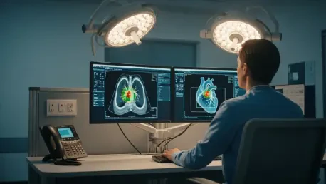
The primary function of this newly cleared technology is to identify and categorize critical positive findings across six primary anatomical regions of interest, including the lungs, pleura, heart, and skeletal structures. Unlike traditional diagnostic software that often operates on a simple binary alert system, this advanced tool provides visual localization through specific bounding boxes that highlight the exact area of concern for the reading radiologist. This approach to explainable artificial intelligence ensures that the human element of medicine remains central to the process, as the software acts as a secondary set of eyes rather than a black-box decision maker. By focusing on these six specific regions, the tool effectively covers the vast majority of findings encountered during routine chest imaging. This comprehensive coverage is essential for detecting subtle abnormalities that might otherwise be overlooked during a busy shift, such as small lung nodules or minor pleural effusions, which often serve as the first indicators of more serious underlying conditions that require immediate intervention.
Furthermore, the integration of these detection capabilities into existing clinical workflows allows for a more streamlined approach to patient management in outpatient clinics and emergency departments. When a potential pathology is identified, the system prioritizes the case within the radiologist’s worklist, ensuring that life-threatening or urgent findings are reviewed before routine screenings. This prioritization is particularly vital for identifying hardware issues or bone fractures that might be secondary to a patient’s primary complaint but are equally important for a holistic treatment plan. Experts in the field emphasize that the early recognition of such findings through automated assistance can lead to a significant stage shift in how diseases like lung cancer are managed. Rather than waiting for symptoms to become severe, the use of this detection tool allows for earlier diagnostic follow-up, which historically correlates with much higher survival rates and less invasive treatment options for patients. This creates a more proactive healthcare environment focused on early detection.
Navigating Regulatory Innovation with Predetermined Change Control Plans
A standout technical and regulatory feature of this recent FDA clearance is the inclusion of a Predetermined Change Control Plan, which distinguishes this software from other chest X-ray detection devices currently on the market. This framework allows the manufacturer to update the underlying AI models and architectures as technology evolves without the necessity of submitting an entirely new regulatory application for every minor iteration or algorithm refinement. This regulatory flexibility is crucial in the fast-paced field of machine learning, where data quality and processing power improve almost monthly. For U.S. healthcare providers, this means they can maintain access to the most advanced versions of the detection algorithm between 2026 and 2028 and beyond, ensuring that the tool remains effective against shifting demographic data or new imaging hardware. This proactive regulatory strategy reflects a sophisticated understanding of software lifecycles, ensuring that the diagnostic tools do not become stagnant or outdated while waiting for traditional bureaucratic approvals.
Beyond the logistical benefits of the change control plan, this clearance highlights a broader industry trend toward more robust and transparent validation processes for medical software. The clinical validation of the tool involved rigorous performance testing, including a multi-reader multi-case study that confirmed the effectiveness of the software as both a standalone detection solution and a supportive aid for seasoned clinicians. By meeting these high standards, the developer has demonstrated that its technology can perform reliably across diverse patient populations and varying image qualities. This level of scrutiny is necessary to build trust among the medical community, which has occasionally been skeptical of artificial intelligence due to concerns about accuracy and reliability. The success of this regulatory submission suggests that the industry is moving toward a standard where AI tools are expected to provide not only high sensitivity but also a high degree of specificity, minimizing the risk of false positives that can lead to unnecessary follow-up procedures and increased patient anxiety.
Strategic Integration and Future Implications for Clinical Practice
Healthcare administrators and lead radiologists took the necessary steps to integrate these diagnostic tools into their existing picture archiving and communication systems to maximize the efficiency of their imaging departments. The deployment of the software across global networks proved that a centralized AI strategy could significantly reduce the time spent on manual screening, especially in regions with a shortage of specialized thoracic radiologists. Medical teams focused on establishing clear protocols for how to handle the automated alerts, ensuring that every localized finding was verified by a human expert before a final diagnosis was issued. This collaborative approach between human intelligence and machine learning helped to optimize the diagnostic pipeline, allowing for faster turnaround times for critical results. By treating the software as a sophisticated triage partner, hospitals observed a marked improvement in the consistency of chest X-ray interpretations, regardless of the time of day or the individual clinician’s level of experience with specific rare pathologies.
Looking toward the future of medical imaging, the focus shifted toward utilizing these detection tools as part of a broader population health management strategy. Organizations began to leverage the data gathered by these AI systems to identify trends in community health, such as rising rates of specific pulmonary conditions in certain geographic areas. This forward-thinking use of diagnostic data allowed for more targeted public health interventions and a more efficient allocation of medical resources. For individual practitioners, the next logical step involved the refinement of these tools to include more longitudinal analysis, comparing current scans with historical images to detect subtle changes over time. The successful implementation of this technology served as a blueprint for the adoption of AI in other diagnostic modalities, proving that when regulatory rigor and clinical utility are balanced, the result is a safer and more effective healthcare system. Leaders in the field continued to advocate for the expansion of these tools into primary care settings, where they could serve as a vital first line of defense in the early detection of chronic diseases.