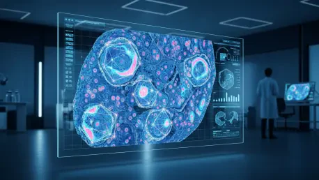

The traditional image of a pathologist peering through a microscope for hours to manually count thousands of stained cells is rapidly becoming a relic of the past as pharmaceutical research demands unprecedented precision. In the high-stakes environment of drug discovery, the transition from qualitative observation to quantitative data is no longer a luxury but a fundamental requirement for success. Modern laboratories are increasingly turning to artificial intelligence to handle the massive influx of data generated by whole-slide imaging systems, which capture tissue samples at a resolution that reveals the most minute biological changes. This shift is driven by the need to identify subtle drug effects that might be invisible to the human eye, particularly when evaluating biomarkers in early-stage trials. By integrating sophisticated algorithms into the workflow, researchers can now analyze tissue architecture with a level of granularity that was previously impossible. This digital transformation does not merely speed up the process; it fundamentally changes the nature of the questions scientists can ask about how a new therapeutic candidate interacts with complex biological systems. As the industry moves toward more complex disease models, the role of AI becomes even more central to maintaining the pace of innovation.
1. Core Advantages: Reliability and Specialized Architectures
The most immediate benefit of implementing AI in preclinical histopathology is the drastic improvement in consistency over traditional human scoring methods. Manual grading is notoriously subjective, with results often varying significantly between different observers or even when the same expert reviews the same slide on a different day. AI-based whole-slide evaluation removes this variability by applying the exact same mathematical parameters across every pixel of a sample, ensuring that biomarker measurements are reliable and reproducible long before clinical trials even begin. This level of precision is critical when measuring low-expression biomarkers where a slight shift in human perception could lead to an incorrect conclusion about a drug’s efficacy. Furthermore, specialized deep learning frameworks have been engineered specifically for the unique challenges of whole-slide images. These architectures, such as those used for nuclear segmentation and multiple instance learning, are designed to process massive file sizes and extract meaningful patterns even when provided with limited data labels. This allows research teams to train highly effective models without requiring thousands of manually annotated images, which has traditionally been a major bottleneck in the development of automated tools.
Beyond simple counting, AI provides standardized safety assessments that are essential for toxicologic pathology and disease-model mapping. When large research groups work together across different geographic locations, maintaining a uniform standard for lesion detection and categorization is a significant challenge. Automated immunohistochemistry scoring minimizes these discrepancies, providing a common language and baseline for all researchers involved in a project. Additionally, modern AI tools offer profound spatial insights by mapping the physical relationships between various cell types within the tissue. While basic cell counts provide a general overview of a sample, architectural context reveals how cells are organized in space, such as the proximity of immune cells to a tumor margin or the distribution of a drug target within a specific organ structure. This spatial data provides a more comprehensive understanding of the biological microenvironment, allowing scientists to see the broader impact of a treatment beyond simple cellular presence. By capturing these complex relationships, AI helps bridge the gap between microscopic observations and macro-level physiological changes, leading to more informed decisions in the drug development pipeline.
2. Research Requirements: Moving Beyond Clinical Categorization
A critical distinction exists between the use of AI in clinical diagnostics and its application within a research-focused preclinical setting. In a clinical environment, the primary goal is often to provide a categorical or binary diagnosis, such as determining whether a tissue sample is malignant or benign. However, research pathology demands a far more nuanced approach, focusing on continuous and precise measurements of biological changes rather than simple classification. Scientists need to know exactly how much a biomarker expression has changed in response to a specific dose or how the density of a particular cell population fluctuates over time. This quantitative focus requires AI models that are tuned for sensitivity and precision across a wide dynamic range of intensities. The ability to generate high-resolution, continuous data allows researchers to construct detailed dose-response curves and perform sophisticated statistical analyses that would be impossible with the discrete categories used in standard medical diagnostics. Consequently, the development of AI for research necessitates a different validation strategy that emphasizes numerical accuracy and the ability to detect minute shifts in tissue composition.
The sheer volume of data generated during preclinical studies also sets research pathology apart, necessitating a level of scale that manual review cannot sustain. A single study can produce hundreds of “gigapixel” slides, each containing billions of pixels of information that must be meticulously examined for subtle signs of toxicity or therapeutic effect. Manually reviewing this volume of material is not only time-consuming but also introduces a high risk of fatigue-related errors and inconsistent findings. AI manages this massive workload by processing images at high speed, maintaining the same level of scrutiny from the first slide to the last. This automated approach allows pathologists to shift their focus from the tedious task of counting cells to the more complex work of interpreting the results and integrating findings into the broader study context. By handling the heavy lifting of data processing, AI ensures that the scale of a study is limited by scientific necessity rather than the availability of human resources. This capability is particularly vital in 2026, where the integration of multi-omics data and high-throughput screening requires histopathology to keep pace with other rapidly evolving fields of drug discovery.
3. Deep Learning Frameworks: Specialized Architectures for Tissue Analysis
To achieve high-quality results in tissue quantification, researchers employ several specialized deep learning frameworks that address specific biological challenges. One of the most prominent is nuclear segmentation, with models like HoVer-Net leading the way in accuracy. These networks are specifically designed to separate crowded or overlapping nuclei, a common problem in dense tissues where individual cells can be difficult to distinguish. By calculating horizontal and vertical distances to the center of each cell, these models can identify individual boundaries with high precision, ensuring that cell counts are not skewed by clusters. This level of detail is essential for accurate biomarker quantification, as it allows for the measurement of expression levels on a per-cell basis. Without such sophisticated segmentation, the risk of miscounting or misidentifying cell types increases significantly, potentially leading to flawed data. These frameworks have become the backbone of modern digital pathology, providing the raw data needed for more advanced downstream analyses and ensuring that the fundamental building blocks of tissue analysis are sound.
Another powerful framework used in research is Multiple Instance Learning, which is particularly effective for analyzing whole-slide images without requiring pixel-level annotations. In this approach, a slide is treated as a collection of smaller patches or “instances,” and the model learns to identify which specific areas are most influential in determining the overall classification or score of the slide. This is incredibly useful in preclinical research where researchers may know the overall status of a tissue—such as whether it came from a treated or untreated group—but do not have the resources to manually label every relevant feature on every slide. MIL allows the algorithm to discover the most relevant biological patterns autonomously, often identifying features that were not previously recognized by human experts. Furthermore, generalist segmentation models like Cellpose have gained popularity due to their versatility. Trained on incredibly diverse datasets, these models can identify cells across a wide variety of tissue types and staining methods without the need for custom retraining. This flexibility makes them an ideal starting point for many research projects, allowing for rapid deployment and providing a reliable baseline for more specialized analysis.
4. Essential Software Tools: Balancing Open-Source and Commercial Solutions
The practical application of AI in the lab relies on a suite of powerful software tools that cater to different research needs and regulatory requirements. QuPath has emerged as a leading open-source platform, widely used for viewing whole slides, detecting cells, and scoring biomarkers across the global research community. Its accessibility and robust feature set allow scientists to experiment with different analysis workflows and share their findings easily. Because it is open-source, QuPath benefits from a large community of developers who constantly contribute new scripts and plugins, keeping the tool at the cutting edge of digital pathology. Researchers often use it as a primary platform for initial exploration and for developing custom analysis pipelines that can be tailored to specific biological questions. Its ability to integrate with other tools and languages, such as Python or R, makes it a versatile hub for complex data science projects within the histopathology lab. This collaborative environment fosters innovation and ensures that even labs with limited budgets can access high-quality analysis tools.
For research conducted in more regulated environments, such as those requiring target validation for pharmaceutical development, commercial platforms like HALO AI provide a validated and streamlined alternative. These systems offer a high degree of user-friendliness and are often preferred for large-scale studies where consistency and audit trails are paramount. Commercial tools typically come with dedicated support and pre-built modules for common tasks like tissue classification or membrane scoring, reducing the time required to set up a new project. In addition to these comprehensive platforms, specialized tools like StarDist are frequently employed for specific challenges, such as segmenting nuclei in regions where they are extremely densely packed. StarDist uses a unique star-convex polygon approach to define cell shapes, which is particularly effective for irregular or crowded biological structures. Meanwhile, Cellpose continues to be a go-to model for broad applications, providing a reliable way to identify nuclei within intact tissue sections across various imaging modalities. The choice between these tools often depends on the specific goals of the study, the complexity of the tissue, and the level of technical expertise available within the research team.
5. Implementation Strategy: A Rigorous Framework for Quantification
Successfully integrating AI into a histopathology workflow requires a structured approach to ensure that the resulting data is both accurate and scientifically sound. The first step in this process is to define a clear reference standard, which involves having one or more experienced pathologists manually mark a representative set of slides. This baseline serves as the “ground truth” and establishes the expected range and appearance of the biomarkers being studied. Without this human-guided foundation, it is impossible to objectively measure how well the AI model is performing. Once the reference standard is established, the next phase is to set up the analysis model by configuring the segmentation and classification software using the marked slides. A crucial part of this step is ensuring that a portion of the images remains hidden from the training process. This “hold-out” set is essential for testing the model’s ability to generalize to new, unseen data, preventing the algorithm from simply memorizing the specific slides it was trained on and ensuring its utility across the entire study.
After the initial setup, the research team must rigorously evaluate performance by measuring the model’s numerical results against the hidden manual markings. This comparison ensures that the AI is accurate across all levels of biomarker intensity, from very low to very high expression. If the model meets the required accuracy thresholds, the full study can be executed, running the verified model on the entire collection of slides using uniform settings. During this phase, it is important to implement a system for flagging any images with technical flaws—such as poor staining, tissue folds, or scanning artifacts—for human review to prevent erroneous data from entering the final analysis. Finally, the implementation framework includes performing regular audits to account for potential shifts in staining quality or scanner settings over time. By routinely checking the model’s performance against small manual samples, researchers can maintain the integrity of their data throughout long-term studies. This disciplined approach ensures that the transition to automated analysis does not come at the expense of scientific rigor, providing a stable foundation for drug discovery programs.
6. Transformation of Target Validation: Integrating AI into Laboratory Workflows
The implementation of artificial intelligence in preclinical histopathology transformed the way research laboratories approached target validation and drug safety. By 2026, the reliance on subjective manual scoring had diminished significantly as digital tools provided a stable, scalable method for analyzing complex tissue structures. Pathologists transitioned into a role where they supervised algorithmic outputs rather than performing the tedious labor of manual quantification, which allowed them to focus on high-level biological interpretation. The integration of these technologies removed the inconsistency inherent in traditional methods, ensuring that drug discovery programs were based on reproducible and transparent data. Researchers successfully utilized deep learning to identify subtle morphological changes that previously went unnoticed, leading to more accurate assessments of therapeutic potential. This shift not only increased the speed of data generation but also improved the reliability of the evidence used to advance candidates through the developmental pipeline, ultimately reducing the risk of failure in later clinical stages.
To maximize the benefits of this technological shift, research organizations should prioritize the standardization of digital workflows and the continuous training of staff in computational pathology. It was established that the most effective labs were those that treated AI not as a standalone solution, but as a collaborative tool that augmented human expertise. Moving forward, laboratories should invest in robust data management systems to handle the massive volume of whole-slide images and the associated metadata. Establishing clear protocols for model validation and regular auditing is essential to maintain data integrity over time. Furthermore, fostering a culture of cross-disciplinary collaboration between pathologists, data scientists, and biologists will be critical for developing models that are both technically proficient and biologically relevant. By embracing these actionable steps, the research community can continue to refine the precision of preclinical studies, ensuring that the next generation of therapies is supported by the most rigorous and comprehensive tissue analysis possible.