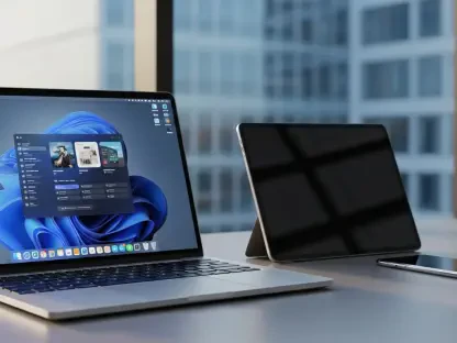

Laurent Giraid is a distinguished technologist who has spent his career at the intersection of artificial intelligence and clinical practice, focusing on how machine learning can enhance surgical precision and ethical safety. As the University of South Florida’s Center for Advanced Medical Learning and Simulation (CAMLS) becomes the first simulation center globally to integrate the Allia Moveo system, Giraid’s perspective is essential for understanding the future of medical education. This conversation explores the shift toward high-fidelity simulation in the Tampa Medical & Research District, the elimination of physical and technical friction in the operating room through cable-free design, and the role of AI in personalizing patient care.
Modern minimally invasive surgery is evolving faster than traditional training models can handle. How does integrating a cable-free, wide-bore imaging system into a simulation environment bridge this gap, and what specific procedural efficiencies should clinicians expect to master before performing live operations?
The traditional “see one, do one, teach one” model is effectively obsolete when you are dealing with the rapid-fire evolution of robotics and AI-enabled workflows. By bringing the Allia Moveo into a simulation center like CAMLS—which is one of the world’s largest free-standing facilities of its kind—we allow clinicians to fail safely while mastering the complex choreography of a hybrid operating room. The cable-free, slim C-arm design is a game-changer because it removes the literal tripping hazards and mechanical limitations that often distract a surgical team during high-stakes maneuvers. Clinicians can now master the “Moveo” mobility, learning to position the imaging system with a fluidity that mirrors the speed of modern minimally invasive surgery (MIS). We expect providers to achieve a level of procedural efficiency where the technology becomes an extension of their own hands, reducing the time spent on equipment adjustment and increasing the time spent on precise clinical execution. This level of training ensures that when the system is installed for clinical use at Tampa General Hospital later in 2026, the staff will already possess the deep muscle memory required for complex cases.
The latest image-guided solutions feature AI-powered workflow tools and slim C-arm designs that allow for unobstructed patient access. Could you walk through a complex procedure where these features reduce technical friction and explain how AI-driven navigation changes the decision-making process for the surgical team?
Consider a complex endovascular repair where every millimeter of accuracy determines the long-term success of the intervention. In a standard setup, the bulky nature of traditional C-arms often forces the surgical team into awkward ergonomic positions, creating “technical friction” that can lead to fatigue or minor errors. With a slim, compact design that offers unobstructed patient access, the primary surgeon and the assisting staff can maintain their natural workflow without fighting for space around the pedestal. The AI-powered workflow tools act as a silent digital co-pilot, processing vast amounts of imaging data in real-time to streamline the navigation through the patient’s vasculature. This shifts the decision-making process from a reactive state—where the team is constantly correcting for poor visibility—to a proactive state where the AI highlights the most efficient path for the catheter. By providing a more personalized patient experience through precise data, the team can make high-pressure calls with a level of confidence that was previously impossible.
Performing Cone Beam CT imaging with a patient’s arms down is a unique capability of this new wide-bore technology. What are the clinical advantages of this approach for diverse patient populations, and how does this level of image quality directly impact outcomes in high-stakes hybrid operating rooms?
In the past, the physical constraints of imaging bores often required patients to have their arms raised, which is not only uncomfortable but often impossible for trauma victims or elderly patients with limited mobility. The wide-bore technology in this new system is specifically designed to accommodate a diverse patient population, allowing for high-quality Cone Beam CT (CBCT) imaging even when the patient’s arms are at their side. This capability is vital in a high-stakes hybrid OR because it eliminates the need to choose between patient safety and image clarity; you no longer have to risk moving a fragile patient just to get a clean scan. The resulting image quality is exceptional, providing the “exceptional mobility” promised by the system to ensure that the surgical team has a 360-degree understanding of the anatomy. When you can see the target tissue or the placement of a device with this much clarity, the risk of complications drops significantly, leading to the shortened hospital stays and reduced pain that define successful MIS.
Establishing a dedicated medical and research district requires deep coordination between academic, clinical, and industry partners. How does placing cutting-edge technology in a simulation center before it hits the hospital floor strengthen the local healthcare ecosystem, and what metrics determine the success of such a model?
The Tampa Medical & Research District (TMRD) is a prime example of how a collaborative ecosystem can accelerate the “bench to bedside” timeline. By placing the Allia Moveo at CAMLS—the first institution in Florida and only the third in the world to receive it—we are creating a training-first culture that builds a massive reservoir of local expertise before a single patient is treated. This strengthens the ecosystem by attracting top-tier clinicians and researchers who want to work with the best tools, effectively turning the region into a magnet for life science innovation. We measure success through metrics such as clinical confidence scores, the speed of technology adoption across multidisciplinary teams, and, ultimately, the patient outcome data from TGH. When we see a measurable decrease in procedure times and a corresponding increase in successful outcomes in the hybrid OR, we know the simulation-first model has worked. This synergy between academic excellence and industry innovation ensures that the latest surgical techniques are not just available, but are being utilized at the highest possible standard.
A clinical version of this image-guided system is often installed in hospitals following its debut in training facilities. What is the step-by-step process for transitioning a multidisciplinary team from simulation to live patient care, and how do you ensure radiation safety standards are maintained during this transition?
The transition begins with high-fidelity simulation where the multidisciplinary team—surgeons, nurses, and technologists—practices “worst-case scenario” drills in the CAMLS environment to ensure seamless coordination. The second step involves mastering the imaging-guided navigation and AI tools in a setting that mimics the TGH hybrid OR suite exactly, so the environment feels identical when they switch to live care. Throughout this process, we place a heavy emphasis on radiation safety, using the system’s automated tools to teach teams how to minimize dose exposure for both themselves and the patient without sacrificing image quality. We track the team’s ability to maintain these safety standards in the lab, providing real-time feedback that becomes part of their formal certification. By the time the second system is installed at Tampa General Hospital later in 2026, the transition is less of a “launch” and more of a “graduation,” where every team member is fully prepared for the realities of live patient care.
What is your forecast for image-guided therapy?
I forecast that image-guided therapy will evolve into a fully autonomous, predictive environment where the “smart” operating room anticipates the surgeon’s next move. We are moving away from imaging being a separate, static tool and toward a future where AI-driven navigation and real-time biological feedback are woven into the very fabric of the surgical theater. Within the next decade, the physical barriers between the surgeon and the digital data will vanish, potentially through augmented reality overlays that allow us to see through tissue as if it were transparent. As we continue to refine systems like the Allia Moveo, the “friction” of the hardware will disappear entirely, leaving behind a streamlined, ultra-precise process that makes even the most complex minimally invasive procedures as routine and safe as a standard check-up.