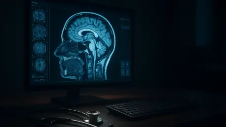

High-performance AI models often suffer from poor generalizability when they are validated on only a single, narrow dataset from one institution. This limitation poses a significant hurdle in neuro-oncology, where the stakes for accurate diagnosis are exceptionally high. In the United States, the five-year survival rate for patients diagnosed with malignant brain tumors is currently 35.7 percent, a statistic that underscores the critical need for early and precise identification of central nervous system malignancies. The diagnostic process is inherently complex because the symptoms of brain tumors, such as persistent headaches, seizures, and cognitive changes, are often non-specific and overlap with many other neurological conditions. Radiologists primarily rely on Magnetic Resonance Imaging (MRI) to detect these lesions, yet the morphological similarities between different tumor types like gliomas and meningiomas can lead to dangerous misclassifications and subsequent errors in treatment plans.
Addressing Critical Gaps in Medical AI
Overcoming Fragmented Workflows: The Black Box Solution
The researchers identified several systemic failures in current medical AI, specifically noting that most models are too specialized to be practical in a fast-paced clinical setting. Many existing systems can either screen for the presence of a tumor or classify its specific type, but they rarely perform both tasks within a single pipeline. This fragmentation forces hospital IT departments to maintain multiple, disconnected software tools, which increases the likelihood of technical errors and slows down the diagnostic process. Furthermore, many high-performing models operate as a “black box,” providing a final diagnosis without explaining which specific visual features of the MRI scan influenced the decision. This lack of transparency is a major barrier to adoption, as clinicians are understandably hesitant to trust a system that cannot justify its conclusions, especially when making life-altering decisions for their patients.
To bridge these significant gaps, the development team engineered a unified and interpretable application that was validated across several large-scale datasets. By focusing on a holistic solution that combines screening and classification, they aimed to create a tool that is not only highly accurate but also transparent and easy to integrate into a radiologist’s daily routine. This approach ensures that the AI serves as a reliable “digital eye” that complements human expertise rather than replacing it with an inscrutable algorithm. The software provides a second opinion that is grounded in data-driven evidence, helping to reduce the cognitive load on specialists who must interpret hundreds of scans every day. By addressing the need for both functionality and clarity, this framework sets a new standard for how artificial intelligence should be implemented within the sensitive domain of modern medical diagnostics.
Architectural Innovation: Dense Connectivity and Robust Design
The system is built on the DenseNet121 architecture, which was strategically chosen for its unique “dense connectivity” where every layer receives inputs from all preceding layers. This specific design allows for extensive feature reuse, which is vital when working with medical datasets that are often much smaller than general-purpose image collections. By ensuring that information flows efficiently through the network, the researchers modified the standard backbone by removing the original top layers and adding a global average pooling layer. These structural changes help the model remain remarkably stable during the training phase and require significantly fewer parameters than other common architectures like ResNet. This efficiency makes the model more robust and less prone to the vanishing gradient problem, which often plagues deep learning systems and prevents them from learning complex patterns in medical imagery during long training cycles.
To prevent the common issue of overfitting, where a model performs well on training data but fails in the real world, the team introduced a custom regularization stack. This includes a dropout layer that randomly deactivates half of the neurons during training, forcing the network to learn redundant and highly reliable features rather than relying on a few specific data points. They also implemented batch normalization to stabilize the internal learning process, allowing the model to converge faster and more consistently across different hardware configurations. These technical refinements ensure that the AI remains accurate when faced with new, unfamiliar data from different medical centers, which has historically been a major stumbling block for medical algorithms. The result is a system that balances complexity with stability, providing a high level of confidence in its diagnostic output across various imaging conditions.
Advanced Methodology and Performance Results
The Dual-Output Strategy: Precision via Specialized Training
A standout feature of this system is its branching output path, which allows it to handle two distinct diagnostic tasks simultaneously. After processing an MRI image through the shared dense layers, the model splits into two directions: one branch determines if a scan is normal or abnormal, while the other classifies the tumor as a glioma, meningioma, or pituitary tumor. To keep the results clean and reliable, the team utilized a clever masking strategy during the training phase. This ensures the model only looks for specific tumor characteristics in scans that have already been identified as abnormal, preventing “noise” from healthy brain images from degrading the accuracy of the final classification. This hierarchical approach mirrors the logical steps a human radiologist takes when reviewing a patient’s file, starting with a general screening before moving into a more detailed and specific pathological analysis.
The performance of this modified model was rigorously tested against several modern architectures, including EfficientNet and Xception, using two massive public datasets. The results were exceptional, with the model achieving a binary screening accuracy of 99.85 percent and a multiclass classification accuracy of 99.00 percent. Most importantly, the system maintained this high performance during “zero-shot” evaluations on completely unseen data from a third institution. This ability to generalize suggests that the AI has successfully learned the fundamental biological features of various tumor types rather than just memorizing the specific pixel patterns from its original training set. This level of cross-dataset reliability is a critical milestone, as it demonstrates that the technology is ready for broader deployment in diverse clinical environments where imaging equipment and patient populations can vary significantly.
Enhancing Usability: From Lab Environment to Clinical Practice
To ensure the model is practically useful for doctors, the researchers integrated Grad-CAM technology, which generates a color-coded heatmap over the original MRI scan. These heatmaps show exactly which areas the AI focused on to reach its conclusion, such as localizing an extra-axial mass or identifying an abnormality in the pituitary region. This visual feedback is crucial for building clinical trust, as it allows a radiologist to verify that the model is looking at the actual pathology rather than background noise or imaging artifacts. Furthermore, the team developed a high-speed desktop application using the PyQt5 framework that provides these comprehensive reports in under 250 milliseconds. This rapid inference time makes the tool fast enough for high-volume hospital environments, where every second saved can lead to faster treatment initiation and better overall management of the patient’s care.
In light of these findings, medical institutions should have prioritized the integration of explainable AI into their standard neuro-radiology protocols. The research demonstrated that a high-performance system could effectively bridge the gap between algorithmic complexity and clinical utility by focusing on transparency. Future efforts were directed toward expanding this dual-task framework to include full pixel-level segmentation and the categorization of tumor grades. Clinicians were encouraged to adopt a human-in-the-loop approach, where AI insights served as a secondary check rather than a final verdict. By establishing a feedback loop where specialists validated model outputs, the medical community ensured the continuous refinement of these digital assistants. This proactive strategy facilitated more personalized treatment plans and ultimately contributed to a measurable improvement in patient outcomes, transforming the standard of care for brain tumor diagnosis.