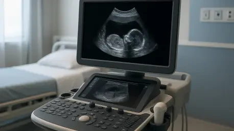

The effectiveness of synthetic medical data is validated when a classifier trained on artificial images maintains high accuracy on entirely unseen, real-world clinical scans. This principle stands at the forefront of modern oncology, where the integration of artificial intelligence is revolutionizing how clinicians detect and treat disease. However, a significant hurdle remains: the scarcity of high-quality, labeled medical data. Training deep learning models requires thousands of expertly annotated images, which are often difficult to obtain due to strict patient privacy laws and the high costs of clinical infrastructure. To overcome this persistent data bottleneck, researchers Ali Hamza and Martin Mezl from the Brno University of Technology have introduced UltraGANet. This framework uses generative AI to synthesize realistic medical imagery, addressing the common problem of overfitting. By utilizing a conditional generative adversarial network, the system creates a robust training ground that allows diagnostic tools to achieve high levels of accuracy without requiring an unattainable volume of real-world patient data.
Harnessing the Power: Generative Adversarial Networks
At the heart of the UltraGANet framework is the Generative Adversarial Network, a dual-network architecture where two AI components compete in a sophisticated digital contest. The first component, known as the generator, creates synthetic grayscale ultrasound images from random mathematical noise, while the second, the discriminator, attempts to distinguish these fakes from real clinical scans. Through this constant competition, the generator eventually learns to produce images so realistic that they are virtually indistinguishable from those taken in a hospital setting. This process is particularly vital for breast ultrasound, a modality favored for its safety and cost-effectiveness but one that often suffers from image noise and variability. By mimicking these complexities, the AI creates a diverse library of training material that prepares diagnostic systems for the unpredictable nature of clinical practice. This method ensures that the resulting diagnostic models are not just memorizing data points but are developing a profound understanding of tissue characteristics.
What makes UltraGANet particularly effective is its conditional nature, which allows researchers to command the AI to produce specific types of images, such as malignant or benign tumors. This capability is vital for balancing datasets, as malignant cases are often rarer in clinical collections than healthy or benign ones. Traditional data augmentation methods, such as rotating or flipping images, offer limited relief because they do not introduce new pathological features. In contrast, the conditional generative adversarial network produces entirely new variations of tumors on demand. By generating a perfectly balanced set of training data, the AI learns to recognize the nuanced features of cancerous tissue with the same proficiency as more common medical findings. This balanced approach eliminates the bias that often plagues medical AI, ensuring that rare but critical pathologies are not overlooked. The ability to synthesize specific classes of disease represents a major shift toward more inclusive and accurate diagnostic tools that can be tailored to various medical needs.
Prioritizing Efficiency: Global Healthcare Deployment
Unlike many modern AI projects that require massive and energy-hungry computing clusters, UltraGANet utilizes a lightweight convolutional neural network as its core classifier. The researchers intentionally avoided the trend of building giant models with hundreds of millions of parameters, opting instead for a compact architecture that requires less memory and processing power. This design choice is grounded in the reality that most medical clinics, particularly those in remote areas, do not have access to high-end supercomputers. By focusing on a streamlined model, the Brno team has ensured that their diagnostic tool can run on the hardware already available in many standard medical environments. This approach prioritizes clinical utility over theoretical complexity, making high-level diagnostic support accessible in a wide variety of settings. The use of such lightweight architectures reduces the barrier to entry for smaller hospitals that wish to implement advanced screening programs without making significant investments in new server infrastructure.
By focusing on computational efficiency, the team has created a system that is ideally suited for real-world deployment in diverse and demanding environments. Portable ultrasound machines in rural or lower-resource areas can benefit from this high-level diagnostic support without needing a high-speed connection to a remote cloud server. This strategy is a prime example of frugal AI, which emphasizes making the benefits of generative technology reach patients in every corner of the globe regardless of local technical resources. Furthermore, the reduced power requirements of these models contribute to a more sustainable implementation of AI in healthcare, minimizing the carbon footprint associated with massive model training. As medical technology continues to move toward the edge of the network, the ability to perform complex image analysis locally on handheld devices becomes a game-changer. This ensures that a patient in a rural community receives the same quality of initial screening as one in a major urban medical center, democratizing access to early detection.
Validating Performance: Rigorous Testing Protocols
To prove that synthetic data can truly train a reliable diagnostic tool, the researchers subjected the UltraGANet framework to a series of stringent evaluations. They utilized five-fold stratified cross-validation to ensure the model’s stability, confirming that its performance was consistent across different segments of data. Following this, the system was tested against a completely independent set of real ultrasound images that the AI had never encountered during its development phases. The results demonstrated that the AI was not just guessing based on its training data but had developed a genuine understanding of breast anatomy and pathology. Specifically, the model achieved a mean accuracy of 91.4% during cross-validation and maintained a high level of performance on the external test set. This validation step is crucial for establishing trust between medical professionals and AI systems, as it demonstrates that the synthetic training process translates effectively to real-world clinical scenarios where the stakes are life-altering.
The performance metrics were particularly impressive regarding clinical safety, showing a 92% recall rate for malignant tumors. In the field of medical diagnostics, a high recall rate is the most critical factor because it indicates the AI is highly unlikely to miss a cancerous growth, which is the most dangerous error a diagnostic tool can make. By maintaining consistent precision across all categories, including benign and normal tissue, the model proved that machine-generated insights could match or even exceed the reliability of traditional training methods. This high level of sensitivity ensures that patients with potential malignancies are correctly flagged for further investigation by a radiologist. The consistency of these results across multiple categories also indicates that the synthetic images provided a comprehensive education for the classifier, teaching it to distinguish subtle differences between various tissue types. This level of reliability suggests that generative AI could become a standard component in the training pipelines for all future diagnostic software.
Ensuring Quality: The Reality of Synthetic Imagery
A major concern with generative AI in medicine is whether fake images maintain the structural integrity necessary for accurate learning. If the generated images look real to a human but lack the underlying textural signatures of a tumor, the resulting diagnostic model would be useless or even dangerous. The researchers addressed this by using structural similarity measures and qualitative assessments to verify that the synthetic scans preserved the essential anatomical features found in original clinical datasets. These measures confirmed that the AI-generated images adhered to the laws of ultrasound physics and reflected the true biological appearance of breast tissue. Interestingly, the study found that a moderate degree of variation in the synthetic images actually helped the model perform better. These machine-generated variations served as edge cases that forced the classifier to become more robust, teaching it to handle the visual noise and irregularities that are common in real-world ultrasound scans but often absent from clean laboratory datasets.
The success of this approach highlights the potential for machine-generated reality to fill the gaps left by the difficulties of human data collection. By teaching the AI to dream up new, anatomically correct variations of tumors, the researchers created a more resilient system that is less prone to the quirks and biases of small datasets. This successful synthesis of data-centric AI and generative modeling suggests a future where the images used to save lives are, in part, manufactured by the machines themselves. Such a paradigm shift allows for the creation of vast, specialized datasets for rare diseases that would otherwise take decades to compile manually. As the technology matures, the focus will likely shift from simply collecting more data to generating better data. This evolution ensures that AI models are trained on the most challenging and representative examples possible, leading to a significant reduction in diagnostic errors. The use of synthetic imagery thus marks a new era in medical education where both humans and machines learn from a mix of real and virtual experiences.
Future Considerations: Strategic Implementation Steps
The study conducted by the researchers at Brno University of Technology established a significant milestone in the application of generative AI for oncology. Researchers observed that the UltraGANet framework successfully bridged the gap between synthetic data generation and practical clinical classification. This achievement was particularly evident in the high recall rates achieved for malignant cases, which surpassed many traditional models trained on limited real-world datasets. The investigation also confirmed that lightweight architectures could compete with larger models when supplied with high-quality, class-specific data. By demonstrating these results, the study provided a clear path forward for the development of portable, AI-enhanced diagnostic tools. The findings highlighted that the bottleneck of data scarcity could be bypassed through strategic use of adversarial training. These outcomes suggested that the healthcare industry was ready to move beyond the limitations of manual data annotation and toward a more automated and scalable future in medical imaging.
Moving forward, the primary focus must shift toward multi-center validation to ensure these models perform across different ultrasound hardware and diverse patient demographics. Developers should prioritize the integration of these generative frameworks into existing clinical workflows, ensuring that radiologists remain at the center of the diagnostic process as ultimate decision-makers. Regulatory bodies will need to establish new standards for validating synthetic training data to ensure safety and transparency in AI-driven medical devices. Additionally, exploring the application of UltraGANet to other types of cancer imaging, such as MRI or CT scans, could further expand its impact. The long-term success of this technology depended on building trust through continued transparent research and real-world pilot programs. By taking these actionable steps, the medical community helped transition generative AI from a promising research concept into a life-saving clinical reality. The journey toward fully automated, high-accuracy diagnostics proved that machine learning could indeed overcome the most daunting data challenges in modern medicine.