
The microscopic interior of a human cell resembles a hyper-connected metropolis where millions of molecular conversations happen simultaneously, yet traditional analytical tools often fail to capture the full story of how these interactions lead to health or disease. Scientists currently navigate a landscape where biological data is more abundant than ever before, yet this wealth of information often obscures the very mechanisms it is intended to reveal. While one scientific instrument captures the structural layout of cellular components, another records the electrical grid of gene expression, and a third tracks the traffic flow of proteins. The core challenge for modern medicine remains the fact that these observations are rarely integrated into a single narrative. We see the isolated parts, but the unified state of the cell—and the precise moment it slips into a pathological condition—remains blurred by fragmented and disconnected data streams.
The Modern Biological Bottleneck: Data Without Clarity
The current era of biological research is defined by a massive influx of information that lacks a corresponding level of interpretability. Every cell functions as a singular, integrated unit, but the methods used to study them often slice that reality into narrow, artificial categories. This fragmentation creates a bottleneck where researchers possess the “ingredients” of cellular activity but lack the “recipe” that explains how they combine to drive systemic behavior. Because the various layers of a cell are inherently linked, much of the data produced by modern sequencing and imaging is redundant. A genetic trigger might manifest as both an invisible RNA sequence and a visible change in the cell’s physical shape, yet standard analysis often treats these as unrelated events.
Without a sophisticated method to separate what is unique to a specific measurement from what is shared across the entire system, the underlying regulatory mechanisms remain hidden. This lack of clarity is particularly problematic when studying the transition from a healthy state to a diseased one. Scientists struggle to pinpoint which specific biological lever was pulled to initiate a tumor’s growth or a neuron’s decay. The result is a critical gap in the ability to design targeted therapies that address the root causes of illness rather than just managing the downstream symptoms. To advance, the field requires a move away from simply collecting more data toward a focus on distilling meaningful insights from the noise of overlapping biological signals.
The Quest for a Holistic View of Cellular Health
Understanding complex conditions like cancer, Alzheimer’s, or diabetes demands a shift toward a comprehensive perspective of how different biological layers interact in real time. Currently, researchers rely on specialized modalities—techniques used to measure transcriptomics, proteomics, or morphology—to gather information. However, the inherent connectivity of cellular systems means that looking at one modality in isolation provides only a partial truth. If a researcher only examines the DNA structure, they might miss the subtle protein changes that actually trigger a disease. The goal is to create a holistic view that acknowledges the cell as a single biological entity where every part influences the whole.
This quest for a unified understanding is hampered by the fact that high-resolution snapshots are often static and disconnected. To truly map disease states, medicine needs to see the “bigger picture” of how physical signals and chemical triggers work in tandem. For instance, in a diseased state, the way a cell moves or changes shape is often a direct consequence of its internal genetic programming. By bridging the gap between these different perspectives, it becomes possible to identify the specific regulatory pathways that drive disease progression. This integrated approach is essential for the development of precision medicine, where treatments are tailored to the unique cellular landscape of an individual patient.
Beyond the Black Box: Disentangling Multimodal Information
To address these challenges, a collaborative team from the Broad Institute, ETH Zurich, and the Paul Scherrer Institute has developed an AI framework that moves past the limitations of traditional models. Standard machine learning tools, such as autoencoders, typically compress all available cellular data into a single, “black box” representation. While this approach is computationally efficient, it “lumps” data together in a way that makes it impossible for researchers to backtrack and identify which specific cellular component is responsible for a particular signal. This lack of transparency prevents scientists from understanding the individual roles of DNA, RNA, or proteins in the broader context of cellular health.
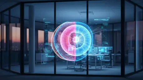
The new framework functions as a sophisticated sorting mechanism that utilizes a unique architecture of shared and modality-specific spaces. This can be compared to a biological Venn diagram where the shared space identifies the core behavior of the cell that is common across all measurements. Meanwhile, the modality-specific spaces isolate data points that are unique to a single technique, such as specialized imaging or sequencing details. This separation allows for a much higher degree of interpretability, ensuring that researchers can finally see both the “forest” of the overall cell state and the “trees” of specific molecular markers.
The framework relies on a specialized two-step training procedure designed to handle the high-dimensional complexity of biological data. This method allows the model to accurately distinguish between overlapping signals and unique markers, maintaining high precision even when analyzing entirely new patient datasets. By training the AI to recognize these distinct layers, the research team has created a tool that can maintain a high level of precision while providing a clear roadmap of how different cellular components regulate one another. This move away from “data lumping” represents a fundamental shift in how artificial intelligence is applied to the life sciences.
Validation Through Real-World Clinical Insights
The true potential of this framework has been demonstrated through its ability to turn abstract data into actionable clinical insights. In validation tests, the AI successfully distinguished between gene activity and DNA structure, identifying which specific markers were most relevant to disease progression. Most notably, the tool was used to identify protein markers associated with DNA damage in cancer patients. By determining exactly which modality—whether it was high-tech sequencing or traditional imaging—captured these damage markers most effectively, the AI helps clinicians choose the most efficient tools for tracking a disease over time.
Led by experts Xinyi Zhang and Caroline Uhler, the research proves that disentangling data provides a clearer lens than traditional integration. Expert analysis confirms that this method allows for a more nuanced interpretation of how a cell reacts to physical signals versus chemical triggers. This level of detail was previously obscured by data overlap, but the new AI framework brings these interactions into sharp focus. The results suggest that by stripping away the noise of redundant information, scientists can uncover the specific biological drivers that were once hidden in the complexity of multimodal datasets.
Strategic Applications in Personalized Medicine and Research
The ability to map the underlying cell state provides a foundation for more efficient and predictive medical research. By analyzing the interaction between a cell’s physical shape and its internal gene expression, researchers can better predict how a tumor might respond to a specific drug or how a neuron might degrade in the early stages of neurodegeneration. This predictive power allows for the development of treatments that address the functional state of the cell, offering a more robust approach to personalized medicine. Instead of a one-size-fits-all treatment, therapies can be adjusted based on the specific “disentangled” profile of a patient’s cellular health.
Furthermore, this AI framework serves as a strategic tool for optimizing experimental design and reducing costs. Measuring every single aspect of a cell is prohibitively expensive and time-consuming, but the model can help scientists determine which measurements are essential and which can be accurately inferred from existing data. This approach streamlines clinical trials and accelerates the pace of discovery by focusing resources on the most informative biological markers. As the field moves toward a unified narrative of human health, this technology removes the guesswork from cellular analysis, allowing for a clearer understanding of the integrated biological systems that sustain life.
The collaborative development of this disentanglement framework successfully addressed the persistent issue of data overlap in single-cell analysis. Researchers utilized the model to bridge the gap between transcriptomics and imaging, resulting in a more precise identification of the regulatory pathways involved in cancer progression. By moving beyond the “black box” limitations of standard autoencoders, the team established a new standard for transparency in biological AI. This shift allowed for the prediction of cellular responses to external stressors with unprecedented accuracy, proving that the separation of shared and specific data is the key to unlocking the mysteries of the cell. Looking forward, the application of this framework will likely expand toward the early detection of metabolic disorders and the refinement of gene editing techniques, ensuring that the next generation of medical interventions is guided by a clear and integrated view of the cellular world.