
In the ever-evolving world of medical advancements, artificial intelligence (AI) has been at the forefront of revolutionizing various fields, and the realm of ophthalmology is no exception. Among the most notable progressions in this domain is the extraordinary collaboration between electrical engineers and ophthalmologists at the University of California, San Diego. Combining expertise from the Jacobs School of Engineering and the Jacobs Retina Center at Shiley Eye Institute, this partnership has paved the way for groundbreaking developments in diagnosing and treating retinal diseases through sophisticated AI and image processing tools.
Interdisciplinary Collaboration for Better Patient Outcomes
Leveraging AI to enhance the capabilities and decision-making processes of ophthalmologists has been the cornerstone of this collaboration. The ultimate goal is to achieve better outcomes for patients suffering from retinal diseases, such as macular degeneration and diabetes-related blindness. Under the guidance of Professor Truong Nguyen, electrical engineering graduate students spend one day each week at the Jacobs Retina Center. Although they do not engage in patient treatment, their role involves working alongside ophthalmology experts to address both clinical and computational challenges.
Over the past five years, this partnership has yielded impressive results, with 21 papers published, contributing to advancements in both clinical understanding and engineering techniques. Key research areas include improving computer vision and AI tools, enhancing image processing methods, predicting the efficacy of specific drugs for individual patients, and aiding in developing new therapeutic treatments for retinal diseases. This interdisciplinary approach has proven essential in identifying innovative solutions to tackle pressing healthcare challenges, showcasing the vast potential of collaborative efforts.
Breakthroughs in AI-Driven Diagnosis
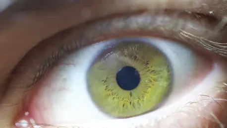
One of the most significant achievements from this collaboration is the development of an AI tool capable of predicting age-related macular degeneration by analyzing optical coherence tomography (OCT) angiography images of eye blood vessels. These noninvasive images can be obtained during a standard clinical visit within minutes, allowing the AI tool to match and even surpass human experts in accuracy, achieving an 80% success rate based on images alone. This remarkable development facilitates faster and more precise diagnoses, ultimately leading to the creation of more effective drugs for treating macular degeneration.
Furthermore, a notable method has been devised to synthesize multiple images taken over time to accurately determine any growth in blood vessel damage or tumors. This process involves overlapping previous images with current ones, enabling quick and precise assessments of changes. The AI method developed for this purpose localizes areas of concern 37% faster than traditional side-by-side comparisons while completely eliminating the error rate associated with human ophthalmologists’ visual checks. These advancements underscore the potential of AI to dramatically improve diagnostic accuracy and efficiency.
Enhancing Image Processing and Predictive Analytics
The research team has also made significant strides in enhancing image processing and predictive analytics. Deep learning networks have been developed to correct eye motion in 3D retinal imaging and precisely quantify morphological changes in vasculature due to age-related macular degeneration using OCTA angiography. Additionally, they have devised methods to correct distortions between ultra-widefield and narrow-angle retinal images and created AI tools to synchronize multimodal images from different optical instruments in patients with retinitis pigmentosa. Such advances highlight the transformative power of AI in augmenting the precision and effectiveness of clinical ophthalmology, ensuring more accurate results and better patient care.
Professor Nguyen underscores the crucial nature of interdisciplinary collaboration, blending clinical insights with advanced machine learning and image processing expertise. This synergy has been instrumental in achieving successful outcomes and significantly impacting both engineering and medical fields. The partnership between Nguyen and William Freeman, MD, Distinguished Professor and vice chair of the Viterbi Family Department of Ophthalmology, exemplifies the rarity of sustained interdisciplinary collaborations. Engineers actively engaging with patient care issues foster a deeper understanding of both fields’ work, leading to remarkable breakthroughs.
Real-World Impact and Future Directions
In the ever-evolving landscape of medical progress, artificial intelligence (AI) stands out as a key driver of change across numerous fields, with ophthalmology being a prime example. One of the most impressive advancements in this area is the remarkable partnership between electrical engineers and ophthalmologists at the University of California, San Diego. This collaborative effort, which merges the expertise of professionals from the Jacobs School of Engineering and the Jacobs Retina Center at Shiley Eye Institute, has spearheaded groundbreaking breakthroughs in the diagnosis and treatment of retinal diseases. By leveraging sophisticated AI and advanced image processing tools, this team has made significant strides in improving retinal health. These innovative technologies not only enhance the accuracy of diagnosing retinal conditions but also boost the effectiveness of treatment plans, offering new hope for patients. This cutting-edge collaboration signifies a remarkable integration of engineering prowess and medical expertise, marking a significant milestone in the field of ophthalmology.