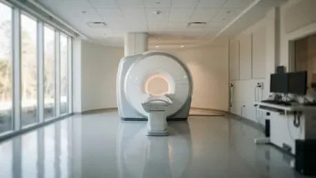

Ischemic stroke remains a leading cause of global mortality, placing immense pressure on clinical teams to identify infarcted brain tissue rapidly enough to determine the viability of reperfusion therapies. In the high-stakes environment of an emergency department, every second counts, and the primary diagnostic hurdle often involves the use of intravenous contrast agents. While these chemical dyes are effective at highlighting damaged vasculature in Magnetic Resonance Imaging (MRI), they present significant risks for patients with chronic kidney disease or acute renal failure. Furthermore, the time required to administer contrast can delay critical intervention, making it a less-than-ideal solution for streamlined triage. Engineers at the Universidad Politécnica de Cartagena have identified this bottleneck and shifted their focus toward non-contrast imaging, specifically utilizing Fluid-Attenuated Inversion Recovery (FLAIR) sequences to improve stroke lesion identification through artificial intelligence.
Technical Challenges in FLAIR Imaging
The transition to non-contrast-based stroke detection is significantly hampered by the inherent visual limitations of Fluid-Attenuated Inversion Recovery sequences. While FLAIR is universally available on clinical scanners and excellent for visualizing tissue swelling, automated segmentation in these images is notoriously difficult because stroke lesions often manifest as faint or diffuse patches rather than the bright, high-contrast spots seen with chemical enhancement. Without the aid of contrast, the artificial intelligence must rely on subtle textural changes and signal intensity shifts that are often difficult to distinguish from healthy brain tissue. This lack of clear boundaries presents a fundamental engineering challenge, requiring the development of algorithms that can identify pathological anomalies in a noisy environment where the difference between a life-threatening lesion and standard tissue is measured in minute gradations of gray.
Beyond the subtle appearance of lesions, several confounding biological and technical factors further complicate the detection process for automated systems. Acute stroke lesions frequently mirror age-related changes known as white matter hyperintensities, which are benign physiological alterations common in older populations. An AI model that is not sufficiently robust may struggle to differentiate these chronic, age-related marks from a sudden, acute infarction, leading to potentially dangerous false positives. Furthermore, inconsistencies in image quality between different hardware manufacturers—such as variations in magnet strength or signal-to-noise ratios—introduce technical artifacts that can obscure pathological findings. To address these nuances, modern deep-learning models must incorporate sophisticated spatial analysis techniques that allow them to filter out physiological variation and focus strictly on the specific morphological characteristics of vascular damage.
Evaluating Diverse Deep Learning Architectures
To investigate the viability of automated stroke screening without contrast, researchers utilized the ISLES 2022 dataset, a comprehensive collection of 250 stroke cases validated by expert radiologists. The study benchmarked three distinct engineering frameworks: the self-configuring nnU-Net, the high-speed YOLOv8 architecture, and a traditional 3D U-Net baseline. The nnU-Net framework is particularly significant because it automatically adjusts its internal architecture based on the specific properties of the input data, effectively removing the need for manual hyperparameter tuning. In contrast, the YOLOv8 model was adapted from real-time object detection tasks to process 3D brain volumes by reconstructing individual 2D slices into a cohesive volumetric output. By training these models exclusively on FLAIR sequences, the team ensured that the systems could not rely on other imaging modalities, such as diffusion-weighted imaging, to identify the location and extent of the stroke lesions.
The performance analysis revealed that modern architectures significantly outperform legacy systems, with the nnU-Net framework achieving the highest Dice similarity coefficient of 0.483. This score represented a substantial 64% improvement over the traditional 3D U-Net, illustrating how much medical imaging technology has progressed in recent years. One of the most intriguing findings of the research was the performance of YOLOv8, which achieved a Dice score of 0.476, trailing the state-of-the-art nnU-Net by a negligible margin. This suggests that lightweight architectures designed for high-speed object detection are becoming increasingly viable for complex medical applications, offering a potential pathway toward real-time clinical assistants. However, despite these advancements, the absolute scores remain in a moderate range, indicating that while AI can identify obvious damage, it is not yet capable of matching the precision of a veteran radiologist in mapping complex lesion boundaries.
Detection Volatility and Clinical Constraints
A critical observation from the study involved the volatility of the detection results across different case profiles. Even the most advanced models occasionally failed to detect any lesion at all in specific test cases, particularly when the stroke damage was exceptionally small or located in areas with minimal contrast against the surrounding white matter. This all-or-nothing phenomenon highlights a significant gap in current deep-learning logic: the systems are proficient at identifying large, obvious pathologies but may overlook the subtle visual cues of an early-stage or minor infarction. Qualitative analysis showed that while nnU-Net produced anatomically coherent boundaries for larger lesions, it struggled with the same sensitivity issues as its peers when faced with diffuse or faint edges. This suggests that the current generation of AI is better suited as a preliminary triage tool rather than a definitive diagnostic solution that can work independently of human supervision.
Several systemic barriers remain before these computational models can be fully integrated into hospital workflows and emergency departments. The validation for this research was conducted on a relatively small sample of test cases, which limits the ability to predict how the models will perform across a broader, more diverse patient demographic. In medical AI, true reliability is usually only established after exposure to thousands of varied cases from multiple sources. Furthermore, the absence of external validation—testing the models on data from different medical facilities with varying imaging protocols—means that the systems may be over-fitted to the specific characteristics of the training dataset. Variations in magnetic resonance hardware can introduce subtle changes in image texture that might cause an AI model to fail if it has only been trained on a single manufacturer’s output, necessitating more rigorous multi-center trials in the coming years.
Path Toward Hybrid Diagnostic Solutions
The most promising path forward involved the development of hybrid diagnostic ecosystems that combined the strengths of different architectural approaches. By merging the spatial precision of 3D-oriented models like nnU-Net with the computational efficiency and operational speed of 2D systems like YOLOv8, engineers aimed to create a tool that was both fast enough for acute triage and accurate enough for detailed treatment planning. Such a system utilized high-speed detection to flag potential stroke cases the moment an image was acquired, followed by more intensive volumetric analysis to refine the lesion map. This layered approach minimized the risk of missing small strokes while providing clinicians with the detailed information needed to decide whether to proceed with aggressive reperfusion therapies. This evolution in medical engineering focused on creating a seamless integration between raw data acquisition and actionable clinical insights for time-sensitive emergencies.
Ultimately, the goal of these advancements was to establish a dependable and equitable diagnostic standard that served all patients, regardless of their physiological constraints. For individuals who could not tolerate chemical contrast agents due to underlying health conditions, a reliable non-contrast screening program offered a safer alternative that did not compromise the speed of care. Clinicians looked toward these AI systems not as replacements for expertise, but as robust secondary observers that helped mitigate the effects of fatigue during long hospital shifts. As the technology continued to mature through 2026 and 2027, the focus remained on improving the sensitivity of these models to minor infarctions and ensuring they could function across diverse hardware platforms. By prioritizing the development of contrast-free diagnostic tools, the medical community took a vital step toward a future where rapid, accurate stroke detection was accessible to every patient in need.