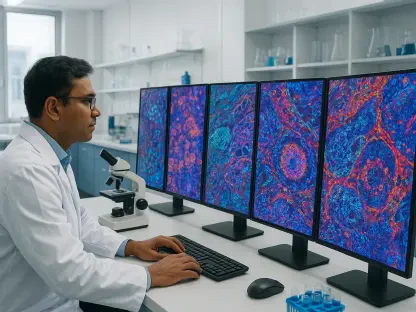

Advanced generative modeling can now steer patches of a light-microscopy image toward their most plausible high-resolution counterparts using optimal transport theory. This breakthrough addresses the central conflict in modern neurobiology, where researchers must choose between the expansive speed of light microscopy and the granular precision of electron microscopy. While the former captures large volumes of brain tissue in hours, it lacks the resolution to map the synaptic connections that define neural circuits. Conversely, electron microscopy provides nanometer-scale detail but proceeds at a pace so slow that imaging a single cubic millimeter of tissue can consume years of effort. The Content-Decoupled Schrödinger Bridge framework emerges as a computational solution to this bottleneck, enabling the recovery of structural information that was previously considered lost to the diffraction limit. This development allows scientists to explore the brain’s wiring with unprecedented efficiency by extracting high-resolution insights directly from standard optical data streams.
The Mathematical Framework: Decoding Stochastic Optimal Transport
At the core of this methodology is the Schrödinger Bridge, a generative model rooted in the principles of stochastic optimal transport. Unlike traditional diffusion models that rely on the gradual removal of noise, the Schrödinger Bridge calculates the most probable path between the blurred data of light microscopy and the sharp detail found in electron micrographs. This mathematical approach ensures that every pixel transformation remains geometrically consistent with the original biological sample, preventing the architectural drift often seen in less sophisticated AI models. By framing the translation as an optimal transport problem, the framework preserves the integrity of the cellular structures while elevating their visual clarity. This shift represents a move away from simple image enhancement toward a principled reconstruction of biological truth based on the underlying statistics of the sample. Consequently, the resulting images are not just clearer versions of the originals but are mathematically rigorous interpretations of the hidden ultrastructural cues within the light signal.
A primary obstacle in applying deep learning to biological imaging is the tendency for models to conflate the physical structure of the tissue with the specific visual artifacts of the microscope. To circumvent this issue, the researchers developed a content-decoupling strategy using a specialized autoencoder that separates modality-invariant content from modality-specific attributes. This allows the system to isolate the actual biological anatomy, such as the shape and position of a neuron, from the contrast and noise patterns unique to light or electron microscopy. Feature-wise Linear Modulation is then employed to inject the necessary high-resolution attributes into the image at precisely the right stage of generation. The result is a synthetic image that possesses the aesthetic and structural characteristics of an electron micrograph while remaining a faithful representation of the specific tissue sample scanned. This decoupling ensures that the generated details are not arbitrary but are strictly tied to the biological content extracted from the optical input data.
Scientific Integrity: Grounding Artificial Intelligence in Physical Laws
The integration of physics-informed constraints serves as a vital safeguard against the common pitfall of AI hallucinations, where models generate convincing but entirely fabricated details. By incorporating the Point Spread Function of the light microscope directly into the learning process, the system is forced to respect the physical limits of how light interacts with lenses. This function describes how a single point of light is blurred by the optical system, and it acts as a rigid boundary for the generative model. Any proposed structural enhancement that contradicts the physical evidence contained within the original optical blur is penalized during the training phase. This approach ensures that the model cannot invent convenient synaptic connections or organelles to satisfy a visual objective; it must find evidence for such structures within the existing data. By grounding the AI in the specific mechanics of the acquisition hardware, the researchers have created a tool that prioritizes scientific accuracy over mere visual improvement.
Further bolstering this integrity is the use of a cross-modality perceptual loss function that maintains the anatomical consistency of the generated images. This loss function evaluates the relationship between the low-resolution input and the high-resolution output, ensuring that the spatial orientation and connectivity of neural processes remain unchanged. This level of rigor is essential for the scientific community, as any deviation from the true biological architecture could lead to incorrect conclusions about brain function or connectivity. The methodology demonstrates that when deep learning is constrained by physical laws and grounded in optimal transport theory, it becomes a much more trustworthy tool for discovery. This shift toward physics-aware models addresses one of the most persistent criticisms of generative AI in the sciences by providing a transparent and verifiable pathway from raw data to enhanced output. As a result, neurobiologists can rely on these computationally derived images with the same confidence they would have in traditional, labor-intensive electron microscopy scans.
Workflow Optimization: Accelerating the Path to Discovery
The practical application of this framework significantly enhances the efficiency of the connectomics pipeline, particularly in the initial phases of data acquisition. In large-scale mapping projects, researchers frequently utilize light microscopy as a scouting tool to identify regions of interest before committing the immense time and hardware resources required for electron microscopy. Because the images enhanced by the Schrödinger Bridge are substantially clearer than raw optical data, human experts can pinpoint critical neural features with far greater accuracy than previously possible. This improved visibility reduces the incidence of missing important landmarks and ensures that expensive electron microscopy time is dedicated only to the most informative tissue samples. By providing a high-fidelity preview of what an electron scan would reveal, the framework allows for a more strategic allocation of resources. This transition from blind scanning to targeted, informed acquisition represents a fundamental shift in how brain-wide data sets are compiled and processed in modern laboratory environments.
Beyond initial acquisition, the framework provides a critical bridge for multi-modal registration, which is the process of aligning images from different types of microscopes. Because light and electron micrographs differ so drastically in appearance, aligning them precisely has historically been a manual and error-prone task. The generated bridge images provide a common visual ground, sharing the anatomical structure of the light microscope with the appearance of the electron microscope, which allows alignment algorithms to function with unprecedented precision. Furthermore, this tool facilitates the use of existing automated segmentation models that were trained exclusively on electron microscopy data. By translating optical data into an electron-like format, researchers can deploy these powerful tracing algorithms on fast light microscopy datasets without the need for the exhausting process of retraining or manually annotating new data. This effectively shifts the bottleneck of connectomics away from the speed of mechanical scanning and toward the efficiency of computational processing.
Collaborative Engineering: Bridging the Divide Between Modalities
The convergence of advanced generative AI and biological imaging represented a broader trend toward computational microscopy, where software became as critical as the hardware lenses. The success of the Content-Decoupled Schrödinger Bridge highlighted that the limitations of optical resolution were not necessarily hard boundaries but rather complex data problems that could be solved through sophisticated modeling. As neurobiology moved toward imaging entire brain volumes in various organisms, the ability to rapidly translate between different imaging modalities proved essential for synthesizing comprehensive maps. This computational bridge allowed for a hybrid approach where the strengths of light and electron microscopy were combined, rather than being treated as separate and incompatible workflows. The economics of brain mapping were increasingly dictated by data throughput, and by moving a portion of this burden from slow hardware to scalable computation, researchers achieved results in weeks that once took years. This development paved the way for more democratized access to high-resolution brain data across the global scientific community.
In summary, the integration of generative modeling with physical constraints established a robust platform for high-speed, high-resolution neurobiological imaging. The researchers demonstrated that the perceived limits of light microscopy were actually opportunities for computational recovery, provided the models remained grounded in the physics of the optical system. Moving forward, the scientific community focused on prioritizing the implementation of these physics-aware frameworks in standard imaging pipelines to maximize the utility of existing hardware. Significant benefits arose from expanding these models to include other imaging modalities, such as fluorescence and super-resolution microscopy, which created a truly universal translation layer for biological data. This proactive adoption of AI-driven resolution enhancement suggested that the future of connectomics resided in the synergy between algorithmic precision and biological observation. By embracing these tools, laboratories drastically accelerated their discovery timelines, which led to a more complete understanding of the neural architectures that underlie cognition and behavior.