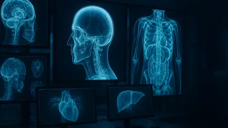

The development of open-source AI tools like Percival encourages global scientific collaboration to refine the accuracy of predictive medical screening technologies. Medical imaging represents a vast, often underutilized reservoir of physiological data that extends far beyond the immediate diagnostic question posed by a treating physician. When a patient undergoes a computed tomography scan to investigate a localized issue such as a suspected bone fracture or acute abdominal pain, the resulting images capture an expansive cross-section of the human body. Hidden within these digital slices are subtle morphological changes and textural patterns that may signal the early onset of systemic diseases long before clinical symptoms manifest. This paradigm, known as opportunistic screening, leverages existing healthcare data to provide additional value without necessitating further radiation exposure or financial costs for the patient. Researchers at the University of Pennsylvania School of Medicine have pioneered this approach by creating a new model.
Data Foundations: Training AI on Longitudinal Medical Records
The foundation of this technological leap lies in the massive scale of information utilized during the development phase of the Percival model. Utilizing the Penn Medicine BioBank, researchers accessed a curated dataset comprising approximately 400,000 CT scans from 50,000 individual patients, providing a diverse representative sample of anatomical variations. Unlike previous iterations of medical AI that were trained on narrow, highly specific datasets to identify a single pathology, this model was exposed to a vast spectrum of imaging data. This breadth allowed the system to learn how to identify complex features across different organ systems simultaneously. By analyzing such a high volume of images, the AI developed a nuanced understanding of what constitutes a normal baseline versus the subtle deviations indicative of underlying health risks. This extensive training was crucial for ensuring that the model could handle the noise and variability found in real-world clinical settings efficiently.
Beyond the raw visual data of the scans themselves, the integration of longitudinal health records played a pivotal role in refining the predictive capabilities of the system. The training process incorporated twenty years of clinical history, including laboratory results, physiological measurements, and documented patient outcomes, allowing the AI to correlate imaging findings with actual disease progression. This holistic approach enables the model to recognize broad relationships between certain imaging patterns and a patient’s long-term health trajectory rather than merely identifying static abnormalities. For instance, the system can connect specific vascular characteristics seen on a chest scan with the subsequent development of cardiovascular complications recorded years later in the patient’s electronic health record. This capability transforms the CT scan from a snapshot in time into a predictive tool that provides insights into a patient’s biological future by learning from these deep temporal connections.
Clinical Validation: Comparing Performance and Future Directions
Rigorous testing was conducted to determine how this new architecture measures up against established automated systems, specifically comparing its performance to the well-known TotalSegmentator tool. The findings indicated that Percival demonstrated superior reliability when flagging complex conditions such as cirrhosis and pancreatic cancer, which are notoriously difficult to detect in their early stages. While traditional tools focus primarily on measuring the volume of specific organs, this model analyzes internal textures and subtle density variations that suggest pathological changes. This enhanced sensitivity is especially valuable for oncological screening, where the difference between a benign variant and a malignant precursor can be minute. However, the researchers emphasize the preclinical nature of this study, noting that the AI is designed to supplement rather than replace the expertise of radiologists. It provides a vital second set of eyes for identifying high-risk markers that might otherwise go unnoted during standard reviews.
The ultimate objective for the research team involved initiating formal clinical trials to measure the tangible impact of AI-driven opportunistic screening on long-term patient health. These studies examined whether early intervention triggered by an AI alert resulted in better management of chronic conditions or improved survival rates for cancer patients. Healthcare administrators explored how to integrate these predictive insights into preventative care models, potentially shifting the focus from reactive treatment to proactive health management. Clinicians were encouraged to view these AI outputs as one component of a broader diagnostic strategy, combining imaging data with traditional physical examinations and genetic risk profiles. By establishing a framework for ethical and practical use, the community prepared for a future where every routine scan became an opportunity for comprehensive assessment. These collective efforts ensured that the technological advancements translated into meaningful changes in global care.