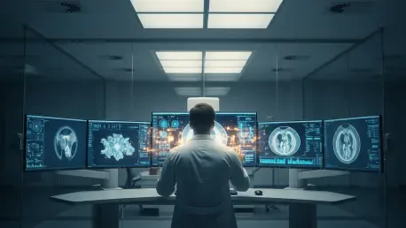

The once-ubiquitous prediction that radiologists would be entirely replaced by automated systems within a few short years has proven to be one of the most significant miscalculations in the history of medical technology. Instead of facing professional extinction, medical imaging specialists are currently experiencing an era of unprecedented growth and integration, where digital tools function not as replacements but as indispensable silicon-based colleagues. This shift has fundamentally redefined the daily workflow within hospitals, moving away from solitary human interpretation toward a sophisticated model of human-machine collaboration that leverages the unique strengths of both biological and artificial intelligence. This evolution highlights a deep-seated misunderstanding of how automation reshapes specialized labor, proving that the human element remains vital even as digital tools become increasingly powerful and pervasive. As healthcare systems globally grapple with rising patient volumes and physician burnout, the emergence of this hybrid model offers a sustainable path forward that prioritizes both diagnostic speed and clinical accuracy. The narrative of competition has been replaced by a pragmatic partnership that addresses the limitations of human perception while grounding machine logic in real-world clinical context. This synergy ensures that every scan is scrutinized with both the tireless precision of an algorithm and the experienced discernment of a medical doctor, creating a standard of care that was previously unattainable.
The Proliferation of AI Tools and Clinical Applications
By the current stages of 2026, the volume of artificial intelligence applications cleared for clinical use has reached a staggering milestone, with approximately three-quarters of the 1,400 medical devices sanctioned by the FDA dedicated specifically to radiological tasks. These tools are no longer experimental novelties confined to academic research; they are operational staples that handle everything from routine administrative report drafting to the high-stakes analysis of complex neurological and oncological imaging. In clinical settings, these systems demonstrate a remarkable capacity to identify subtle abnormalities that the human eye, even one belonging to a seasoned expert, might occasionally overlook during a demanding shift. For instance, recent large-scale clinical trials have validated that AI-assisted colonoscopies consistently identify a higher percentage of polyps compared to traditional manual methods, providing a critical safety net in preventative medicine. Similarly, landmark studies in Sweden have demonstrated that AI-assisted mammography significantly improves cancer detection rates while simultaneously reducing the physical and cognitive workload on radiologists by up to forty percent. These advancements are proving essential in the ongoing effort to reduce global diagnostic error rates, which currently impact millions of patients every year and remain a primary driver for technological adoption in modern healthcare infrastructure across the United States and Europe.
Beyond mere detection, the implementation of these tools is primarily driven by a systemic need for heightened precision and optimized patient outcomes through intelligent triage. By automating the prioritization of urgent cases, such as suspected intracranial hemorrhages or pulmonary embolisms, artificial intelligence ensures that the most life-threatening conditions are flagged for immediate human review the moment the scan is completed. This capability effectively eliminates the traditional “first-in, first-out” queue, which often left critical cases waiting behind routine screenings for hours, potentially worsening patient prognosis. This newfound efficiency allows radiologists to redirect their limited time toward the most complex diagnostic challenges, transforming the speed and accuracy of modern diagnostic workflows across entire hospital networks. Furthermore, the integration of these systems has led to a noticeable reduction in physician fatigue, as the “heavy lifting” of scanning thousands of normal images is increasingly offloaded to digital assistants. This allows the human doctor to arrive at the truly difficult cases with a clearer mind and a more focused perspective, reducing the likelihood of fatigue-related oversights. The result is a healthcare environment where technology serves as an initial filter, ensuring that human expertise is deployed where it can have the greatest impact on patient survival and long-term wellness, creating a more responsive and reliable diagnostic pipeline for all patients.
Developing a Collaborative Framework: The New Medical Standard
The defining challenge for the medical community is no longer a debate about whether to use technology, but rather how to construct a robust framework for an effective partnership. Even as artificial intelligence begins to outperform humans in specific statistical categories, these systems remain prone to unique, non-human errors that a trained physician would never commit, such as misinterpreting a digital artifact or a stray piece of medical equipment as a malignant tumor. Consequently, the radiologist has transitioned into a role of a final judge or “diagnostic gatekeeper,” tasked with reviewing machine-generated insights and deciding when to exercise their critical veto power. This shift requires a significant mental adjustment for physicians who were trained on older, rule-based computer-aided detection systems that followed linear, transparent logic. Modern artificial intelligence relies on complex “black box” neural networks that process millions of data points through layers of calculation that are not easily explained or visualized by a human observer in real-time. This lack of transparency means the radiologist must develop a new kind of professional intuition, learning to interpret not just the patient’s anatomy, but also the reliability of the machine’s suggestions. The collaboration is thus built on a foundation of mutual verification, where the machine provides the raw processing power and the human provides the final layer of clinical validation.
When these high-performance systems identify a subtle lesion or a possible fracture, the physician must determine if the machine has uncovered a genuine clinical concern or if it is simply “hallucinating” a pattern where none exists. This level of oversight is essential to ensure that the increased speed provided by automation does not come at the cost of clinical accuracy or patient safety. The radiologist’s daily activity is becoming less about the rote mechanical task of reading every single image and more about high-level interpretation and the synthesis of disparate data points. This evolving responsibility necessitates a deeper understanding of data science within the medical curriculum, as doctors must now understand the limitations and biases inherent in the algorithms they use. By acting as the ultimate authority in the diagnostic loop, the radiologist ensures that the final report reflects a nuanced understanding of the patient’s condition rather than just a statistical probability generated by a processor. This oversight prevents the “automation of error,” where a mistake made by a machine is blindly accepted and passed through the healthcare system without question. The goal of this partnership is to create a symbiotic relationship where the strengths of the algorithm—consistency and speed—are tempered by the wisdom and skepticism of the human practitioner, resulting in a diagnostic process that is far superior to what either could achieve alone.
Comparing Machine Stamina and Human Context
One of the most significant advantages of artificial intelligence in a clinical setting is its infinite stamina, which allows it to scan every individual pixel of a high-resolution image without the fatigue, eye strain, or distraction that naturally affects humans during a long shift. A machine can compare a single current scan against millions of historical records and reference cases in a matter of seconds, providing a level of comparative data processing that is physically impossible for a person to achieve within a reasonable timeframe. This consistency makes it an invaluable tool for high-volume environments where the sheer number of images to be reviewed can lead to human burnout and an increase in accidental omissions. In contrast, human radiologists provide the essential clinical context and an understanding of a patient’s broader medical history that an algorithm cannot yet fully grasp. While a machine might see a specific pattern of opacities in a vacuum, a human understands the nuances of biology, the patient’s recent surgical history, and the specific environmental factors that might influence a diagnosis. This combination creates a specialized safety net where the human often identifies the specific, outlier cases that the machine misses due to their rarity, while the machine catches the subtle patterns that the human might miss due to exhaustion. This interplay of technology and biology represents the pinnacle of modern medical imaging, providing a dual-layered approach to patient care.
The ultimate objective of this partnership is to establish a workflow where the human remains sufficiently vigilant to dismiss incorrect machine findings while simultaneously remaining open enough to accept accurate ones that might contradict their initial impression. Finding this delicate balance is notoriously difficult because it involves managing complex psychological biases that naturally occur when humans work alongside high-performance technology for extended periods. There is a constant tension between trusting the machine’s superior processing speed and maintaining the professional skepticism required to catch a digital error. Success in this new environment depends on the human’s ability to remain the primary decision-maker in the diagnostic process, using the AI as an advisor rather than a leader. This requires a cultural shift within radiology departments, moving away from a culture of individual autonomy toward one of collaborative intelligence. As these tools become more integrated, the focus is shifting toward how to best present machine data to the human user so that it enhances, rather than overwhelms, their natural decision-making process. By leveraging machine stamina to handle the mundane and repetitive aspects of imaging, the radiologist is freed to engage in the more cerebral and empathetic aspects of medicine, such as communicating complex findings to patients and collaborating with other specialists on personalized treatment plans.
Overcoming Cognitive Obstacles and Training Needs
The daily integration of artificial intelligence into the clinical environment introduces several cognitive risks, most notably automation bias, where a physician might over-rely on a computer’s output and ignore their own professional judgment or visible evidence. Over time, the high reliability of modern systems can lead to a state of automation complacency, where a radiologist might stop looking for the rare but critical errors that the system might commit because it has been correct so many times before. Clinical studies have shown that even highly experienced experts can lose a degree of accuracy when they are presented with incorrect machine predictions, as the social pressure to agree with a “sophisticated” tool can be surprisingly powerful. Conversely, “silly” mistakes made by an artificial intelligence, such as misidentifying shadows caused by minor patient movement or clothing as pathological findings, can lead to a total loss of trust and the eventual rejection of the tool by the medical staff. This knee-jerk reaction is equally damaging to patient care, as it causes physicians to miss the genuine, subtle benefits that the technology offers in the majority of cases. Balancing these two extremes—over-reliance and total distrust—requires a high level of professional maturity and a deep, practical understanding of the specific strengths and limitations of the software being utilized in the clinic, ensuring that technology remains a tool and not a crutch.
Moving forward, the medical field focused on implementing rigorous monitoring protocols and improved educational standards to optimize this evolving partnership between man and machine. Developers were encouraged to provide “confidence estimates” for every finding instead of simple binary answers, which allowed physicians to see exactly how certain the algorithm was about a specific detection. By shifting the radiologist’s role to that of a “diagnostic orchestrator,” the profession ensured that technical precision was always tempered by the depth of human experience and ethical consideration. Hospitals adopted new training modules that specifically taught clinicians how to identify algorithmic failure modes, treating the AI as a trainee that required constant supervision rather than an infallible expert. These steps mitigated the risks of complacency and restored the balance of authority within the diagnostic suite. Furthermore, the industry established clear guidelines for the periodic re-evaluation of AI models to ensure they did not suffer from performance drift over time due to changes in imaging hardware or patient demographics. This proactive approach to technology management solidified the role of the radiologist as the central figure in patient care, utilizing every available digital tool to enhance human capability. Ultimately, the successful integration of these systems transformed the landscape of medical imaging into a more resilient and accurate discipline that benefited patients across the entire spectrum of healthcare, proving that the future of medicine is undeniably collaborative.