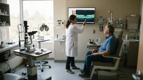

The rigorous use of patient-level stratified splitting during the training process ensures that the AI model can generalize effectively to new, unseen medical data. This foundational approach is critical as the healthcare industry transitions from isolated machine learning experiments to comprehensive, agentic AI frameworks that handle complex clinical workflows. Currently, the field of ophthalmology is grappling with a significant increase in patient volume, particularly regarding conditions like macular holes, which are small breaks in the central retina that can lead to permanent vision loss if not addressed with surgical precision. Traditional AI tools have historically functioned as narrow classifiers, identifying a disease but leaving the heavy lifting of interpretation and patient communication to the human clinician. By 2026, the demand for more autonomous systems has led to the development of integrated pipelines that not only detect pathology via optical coherence tomography but also draft professional clinical reports, effectively bridging the gap between raw data and actionable medical advice. This evolution represents a shift toward technology that acts as a collaborative partner in the clinic rather than just a digital lens, addressing the dual pressure of an aging population and the global shortage of specialized retinal experts. The resulting framework offers a blueprint for more efficient, transparent, and patient-centered eye care.
Structural Evolution: The Modular Architecture
The core innovation of this research lies in its agentic approach, which utilizes a modular design rather than a single, monolithic artificial intelligence. The workflow is divided into three distinct functional layers managed by a central orchestration agent to ensure precision and safety. Instead of building a vision model from scratch, the researchers utilized RETFound, a prominent foundation model for ophthalmology. This model was originally trained on millions of unlabeled retinal images to learn the general architecture of the human eye, allowing the team to fine-tune the OCT-specific branch for the binary classification of macular holes. This specialization ensures that the high-stakes task of medical diagnosis is performed by a tool optimized for visual patterns, rather than a general-purpose text model. By leveraging a foundation model, the system benefits from a deeper understanding of ocular anatomy, which provides a more stable baseline for identifying subtle pathological changes that might be missed by less sophisticated algorithms or generalized computer vision systems.
Following the diagnostic phase, the orchestration agent acts as the brain of the operation, receiving the diagnostic output and deciding how to proceed based on clinical protocols. This component is essential for preventing the language model from hallucinating a diagnosis, as it strictly feeds the vision model’s results alongside standardized medical guidelines into the communication layer. Using GPT-4 via a secured API, the communication engine takes the structured diagnostic data and transforms it into a professional, patient-friendly report. By limiting the role of the large language model to synthesis and formatting, the system avoids the reliability issues often observed when text-based models are asked to interpret medical images directly. This separation of concerns allows for easier updates to the system, as a better vision model or a more advanced language model can be swapped into the workflow without needing to retrain the entire pipeline. This modularity ensures the system remains adaptable in a rapidly evolving technological landscape where medical standards and software capabilities are constantly shifting.
Rigorous Validation: Data Integrity and Reliability
A primary strength of this study is the rigorous management of data to ensure the model generalizes well across diverse clinical settings and hardware. The researchers utilized a variety of datasets, including CHU, OCTDL, and Kermany, to provide a broad training base that reflects different patient demographics and imaging qualities. External validation, which is crucial for proving the model’s efficacy in the real world, was conducted using entirely separate datasets from the OIMHS and Noor Eye Hospital. To prevent data leakage, the team implemented patient-level stratified splitting, ensuring that images from the same individual were never divided between the training and testing phases. This methodological rigor is vital in medical AI to ensure that the performance metrics reflect the model’s actual diagnostic capability rather than its ability to memorize specific image artifacts. By testing against diverse scanners and acquisition protocols, the researchers demonstrated that the agentic framework is capable of maintaining high performance in broad clinical applications beyond controlled laboratory environments.
The results of the study were exceptionally strong, with the model achieving 100% accuracy, sensitivity, and specificity on the internal test set. During external validation involving over 1,000 images, the framework maintained an impressive 98.8% accuracy and 100% specificity. This means the system correctly identified nearly every instance of a macular hole while never incorrectly flagging a healthy eye as diseased. High specificity is particularly important in a clinical setting to avoid unnecessary patient anxiety and unwarranted surgical referrals, which can strain hospital resources and lead to complications. The model’s ability to maintain such high performance across different datasets suggests that the fine-tuned foundation model approach is more robust than traditional deep learning methods. This level of reliability provides a strong argument for the integration of such systems into virtual clinics, where they can act as a high-speed screening tool that identifies critical cases for immediate review by a human ophthalmologist, thereby optimizing the clinical workflow for both doctors and patients.
Visual Evidence: Explainability in Diagnostic AI
To ensure the model was focusing on relevant pathology rather than background noise or imaging artifacts, the team utilized gradient-weighted class activation mapping. These heatmaps provided a visual representation of the areas within the OCT scans that most heavily influenced the AI’s decision-making process. The analysis confirmed that the model’s focus was consistently centered on the retinal break and the surrounding tissue layers, mirroring the diagnostic focus of a human expert. Explainability is a critical requirement for AI adoption in high-stakes medical fields, as clinicians must be able to verify the reasoning behind an automated diagnosis. By providing these visual cues, the framework moves away from the “black box” nature of earlier AI models, offering a level of transparency that builds trust with medical professionals. This feature allows ophthalmologists to quickly confirm that the AI is identifying the correct anatomical features, which is essential for maintaining safety standards and ensuring that the technology assists rather than obscures the diagnostic process.
Statistical tests were also conducted to verify that these visualizations were the result of learned clinical features rather than random architectural artifacts. Through parameter randomization, the researchers proved that the model’s sensitivity to specific retinal structures was deeply rooted in its training rather than coincidence. This statistical validation further strengthens the case for the model’s clinical utility, as it demonstrates a consistent and reliable interpretation of ocular pathology. When compared with the focus of human clinicians, the AI’s heatmaps aligned with the standard markers for macular holes, such as the separation of the sensory retina and the presence of subretinal fluid. This alignment suggests that the fine-tuning process successfully taught the model to prioritize the same clinical indicators that are taught in medical schools. Consequently, the framework does not just provide a result; it provides a clinically relevant justification for that result, which is a necessary step toward the full integration of autonomous diagnostic systems in modern retinal clinics.
Clinical Integration: Automated Reporting and Human Oversight
The transition from a raw diagnosis to a formal clinical report is where many automated systems fail to meet professional standards. In this study, the quality of the automated reporting was evaluated by two independent ophthalmologists who reviewed 40 randomly selected documents generated by the system. The reports were assessed for their diagnostic accuracy and the sufficiency of the management plans and risk disclosures provided to the patient. The results were highly favorable, with 92.5% of the reports rated as both accurate and sufficient for clinical use. This indicates that the AI can mimic the professional tone and content of a human clinician with a high degree of reliability, potentially saving hours of administrative work each week. By automating the drafting of these letters, the system allows doctors to spend more time with patients and less time on documentation, directly addressing one of the leading causes of professional burnout in the healthcare sector.
Despite the high success rate, the reviewers noted that a small percentage of reports lacked certain nuances, emphasizing the importance of a human-in-the-loop verification process. This oversight ensures that the final document sent to a patient meets all professional standards and accounts for any unique clinical variables the AI might have missed. However, the system significantly reduces the time required for clinicians to draft these communications, as the AI-generated draft provides a highly accurate starting point that usually requires only minor adjustments. The integration of large language models for this task demonstrates how generative technology can be safely harnessed when governed by a strict orchestration layer. As medical facilities continue to modernize, these automated reporting tools will likely become a standard feature of electronic health record systems, facilitating faster communication between primary care providers and specialized eye centers. This streamlined approach not only improves clinic efficiency but also ensures that patients receive their results and management plans in a more timely and professional manner.
Evolution: Moving Toward 3D Diagnostics
While the current results are promising, the study acknowledged several hurdles to full-scale clinical adoption that must be addressed in future iterations. The model currently operates on 2D OCT B-scans, whereas real-world clinics often rely on 3D volumetric scans to catch very small or eccentric holes that a single 2D slice might overlook. Additionally, the system was designed for binary classification and does not yet stage the severity of the hole or provide specific prognostic information regarding the likelihood of a successful surgical outcome. Future development will focus on incorporating volumetric analysis and automated image quality assessment, which would allow the AI to reject blurry or low-quality scans and request a retake immediately. This would further enhance the reliability of the system and ensure that only the highest quality data is used for diagnostic decisions. Expanding the system to recognize other retinal conditions, such as age-related macular degeneration or diabetic retinopathy, would also increase its utility as a comprehensive diagnostic tool.
The study concluded that the agentic AI workflow successfully demonstrated the ability to automate the diagnosis and reporting of macular holes with a level of precision that matches human experts. It was observed that the modular architecture provided a safer and more adaptable framework than monolithic models, allowing for a clear separation between image interpretation and linguistic synthesis. The findings suggested that the use of specialized foundation models was the most effective path forward for high-stakes medical tasks. To move toward broader implementation, healthcare providers should consider pilot testing these frameworks in virtual clinic environments where screening volume is highest. The successful validation of this system provided a clear roadmap for the integration of multi-agent AI into daily medical practice, suggesting that the future of ophthalmology lies in the seamless coordination between human expertise and automated intelligence. By focusing on task separation and expert model coordination, the medical community established a robust standard for delivering precise, transparent, and patient-centered care in a digital-first era.