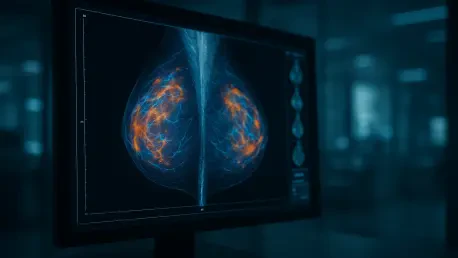

Medical experts caution that simply identifying a tumor on a past scan does not yet prove that real-time AI intervention would have altered the ultimate clinical outcome. The recent analysis by the UCLA Health Jonsson Comprehensive Cancer Center sheds light on the evolving role of algorithmic assistance in breast cancer screening, emphasizing that while high-tech tools are becoming ubiquitous, their true value depends on clinical integration. Radiologists have long struggled with the limitations of traditional mammography, where subtle signs of malignancy can go undetected despite rigorous review. This review serves as a pivotal assessment of how deep-learning models might bridge the gap between initial negative results and the eventual discovery of aggressive interval cancers. By scrutinizing a vast library of historical imaging data, researchers are now determining whether artificial intelligence can truly serve as a dependable second set of eyes in a field where stakes are incredibly high.
Understanding the Clinical Impact: Interval Malignancies
Interval cancers remain a significant challenge because they emerge in the window between scheduled mammograms, often presenting with more aggressive biological profiles than tumors found during routine screenings. These malignancies are generally grouped into two categories: those that were genuinely absent during the last exam due to rapid growth and those known as occult signs, which were present but remained invisible to human interpretation. The research review posits that artificial intelligence excels at identifying these occult markers, potentially offering a window for earlier intervention that was previously unavailable. By training on diverse datasets, these algorithms have demonstrated an ability to recognize morphological nuances that deviate from healthy breast tissue. However, the study notes that the effectiveness of AI varies wildly, with detection rates for future cancers ranging from five to seventy-eight percent. This variation underscores the need for more standardized metrics to ensure that the technology provides consistent benefits.
While retrospective studies show that AI can flag anomalies on past scans that humans missed, translating this capability into a live clinical environment requires a shift in how radiologists interact with automated alerts. The discrepancy between looking backward at known cases and making real-time decisions is a core focus of the analysis. Algorithms may successfully identify patterns in seemingly healthy tissue that suggest a high risk of developing cancer years down the line, with one specific study flagging nearly forty percent of future interval cancer cases. Yet, the presence of an AI flag does not always equate to a clear medical directive. If the machine identifies a risk but there is no visible mass, doctors face a dilemma regarding how to proceed without causing unnecessary patient distress. Building a bridge between these algorithmic insights and tangible patient care requires extensive validation to ensure that earlier detection actually leads to better survival rates and reduced treatments.
Comparative Global Trials: the Hurdle of Standardization
Evidence of the potential of AI is supported by a massive European trial involving more than one hundred thousand women, which evaluated the impact of integrating these tools into a traditional screening workflow. This study compared AI-supported screening against the standard double-reading method where two radiologists independently assess every mammogram. The findings revealed that AI-supported systems achieved higher sensitivity while maintaining a high level of specificity, essentially catching more cancers without significantly increasing false positives. One of the most impactful takeaways from this trial was a reduction in the total screening workload for radiologists by over forty-four percent. This efficiency gain is particularly relevant in a healthcare landscape currently plagued by physician burnout and a shortage of specialized imaging experts. By automating the preliminary review of clear scans, AI allows radiologists to focus their expertise on the most complex and ambiguous cases.
Despite the impressive results from overseas, the investigators caution that the European model may not be a perfect fit for the American healthcare system. The United States primarily utilizes single-physician readings and relies heavily on three-dimensional digital breast tomosynthesis, whereas the European trial was largely based on two-dimensional imaging and a double-reader standard. This difference in baseline infrastructure means that the performance gains observed in one region might not immediately replicate in another. Furthermore, the lack of standardization across the AI industry creates a fragmented landscape where different software developers use varying definitions for what constitutes a high-risk flag or a potential malignancy. Without a unified set of criteria, healthcare providers struggle to compare different products or integrate them into existing workflows efficiently. This non-standardization extends to how data is reported and how results are communicated to patients.
Addressing Overdiagnosis: Establishing Future Clinical Pathways
A primary concern discussed in the review is the risk of overdiagnosis, where AI flags indolent tumors that might never have become life-threatening if left undetected. The introduction of highly sensitive algorithms could lead to a surge in follow-up biopsies and diagnostic imaging for lesions that would have remained asymptomatic throughout a patient’s life. This phenomenon not only adds a financial burden to the healthcare system but also causes significant psychological stress and physical discomfort for patients. There is currently no medical consensus on how to handle high-risk patterns identified by AI when the physical evidence on a mammogram remains inconclusive. Without clear clinical pathways, these technological warnings might lead to aggressive overtreatment rather than watchful waiting or targeted surveillance. The challenge lies in refining models to distinguish between fast-moving, aggressive cancers and slow-growing ones that do not require immediate surgical or chemical intervention.
The UCLA review established that artificial intelligence successfully functioned as a powerful supplementary tool rather than a comprehensive replacement for human expertise. Future research initiatives prioritized post-market surveillance to ensure that these diagnostic algorithms remained effective across diverse populations and varying imaging technologies. Medical institutions moved toward establishing standardized protocols for managing high-risk AI flags, which helped reduce the uncertainty that previously surrounded automated alerts. By focusing on prospective studies that tracked long-term patient survival, researchers gathered the evidence necessary to justify broader clinical adoption. Healthcare providers also emphasized the importance of integrating these tools into a holistic diagnostic framework that balanced technological precision with clinical judgment. Moving forward, the focus shifted to refining these systems to minimize false positives while maximizing the detection of truly life-threatening malignancies.