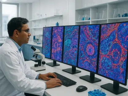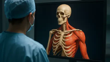

Automating the segmentation of healthy tissues from whole-body scans provides clinicians with a detailed map of a patient’s physical resilience and treatment response. For years, the primary objective of whole-body MRI in the oncology ward was to visualize the extent of malignancy and monitor tumor size. However, researchers at The Royal Marsden NHS Foundation Trust and The Institute of Cancer Research have changed this narrative by introducing an artificial intelligence tool that looks beyond the cancer itself. This software analyzes standard medical imaging to quantify critical biological data such as skeletal muscle mass and fat distribution in patients battling multiple myeloma. By doing so, doctors gain an unprecedented view of how the patient’s body is holding up under the weight of aggressive therapies. This paradigm shift ensures that clinical decisions are based not just on the status of the cancer cells, but on the physiological integrity of the human being.
Revolutionary Precision: The Role of Automated Tissue Analysis
The core innovation of this AI technology lies in its ability to automate the arduous process of tissue segmentation with remarkable speed and accuracy. Previously, if a radiologist wanted to calculate the volume of muscle or fat from a whole-body MRI, they had to manually outline these structures across dozens of imaging slices. This task was so labor-intensive that it remained restricted to research settings and was rarely feasible in a fast-paced clinical environment. Now, the AI algorithm performs these calculations in a matter of minutes, providing a comprehensive report that accompanies standard diagnostic findings. This breakthrough allows for the large-scale application of body composition profiling across patient populations. By removing the bottleneck of manual labor, the tool enables medical teams to monitor physical health markers as frequently as they monitor the cancer itself, without adding to the administrative burden faced by specialists.
Furthermore, the integration of this tool into existing healthcare workflows represents a major leap forward for patient experience and hospital efficiency. Because the AI analyzes standard MRI scans that are already part of the routine care pathway for multiple myeloma, patients do not need to schedule additional appointments or undergo invasive procedures. This non-invasive approach is vital for those undergoing intensive treatments like induction chemotherapy, where reducing the physical and logistical burden is a high priority. The technology effectively turns every diagnostic scan into a multi-dimensional health check, extracting hidden value from data that was already being collected. As hospitals look for ways to optimize resource allocation in 2026, this type of automated data extraction serves as a model for how machine learning can enhance clinical productivity while simultaneously improving the depth of information available to the treating physician.
Strategic Prognosis: Identifying Survival Indicators Through Body Composition
The application of this AI tool has revealed a significant correlation between a patient’s internal physical makeup and their long-term clinical outcomes during treatment. By examining scans at three critical points—prior to treatment, after chemotherapy, and following a transplant—researchers have identified clear patterns in how the body reacts to the rigors of oncology care. One of the most striking findings is that patients entering therapy with higher baseline levels of abdominal muscle and subcutaneous fat demonstrated longer progression-free survival. This suggests that a patient’s physical reserve acts as a vital buffer, allowing them to better withstand the systemic toxicity associated with high-dose chemotherapy. These insights allow clinicians to move beyond a one-size-fits-all approach, recognizing that the internal structural health of the patient is as predictive of success as the genetic profile of the tumor.
Conversely, the AI-driven data highlighted the risks associated with certain types of fat accumulation during the treatment process. Specifically, an increase in visceral fat, which is the tissue surrounding internal organs, was linked to a significantly higher risk of disease progression. While subcutaneous fat may provide a beneficial energy reserve, visceral fat is often associated with inflammation and metabolic dysfunction that can complicate cancer management. The ability to distinguish between these two types of fat in a clinical setting provides doctors with a window into the patient’s internal environment that was previously unavailable. This nuance is critical because it allows for the identification of high-risk physiological shifts that would otherwise go unnoticed until physical symptoms manifested. By monitoring these fat dynamics, medical teams can intervene early, potentially altering the patient’s metabolic trajectory and improving remission chances.
Clinical Integration: The Future of Holistic Cancer Management
The implications for clinical practice extend into the refinement of drug development and the optimization of existing therapeutic combinations. By observing how different drug regimens impact a patient’s physical well-being alongside their efficacy in killing cancer cells, researchers can develop more nuanced treatment protocols. For example, if a specific combination of immunotherapy and chemotherapy is found to cause less muscle wasting than another while maintaining efficacy, it could become the preferred choice for frail patients. This data-driven approach allows for a physical health baseline to be established during trials, ensuring that new treatments are evaluated not just by their ability to extend life, but by their ability to preserve the quality of that life. As this technology becomes standardized, it will enable a level of comparative analysis that was previously impossible, leading to a more effective standard of care for patients who must undergo years of continuous medical intervention.
The validation of this AI-powered imaging tool established a new standard for how medical data could be utilized to enhance complex cancer care. By transforming routine MRI scans into comprehensive maps of physical resilience, the research demonstrated that the fight against multiple myeloma required a deeper understanding of the patient’s physiological landscape. Moving forward, the implementation of this technology across diverse clinical settings will necessitate further trials to ensure these biomarkers are predictive across all demographic groups. Health systems should look toward integrating these automated reports into standard diagnostic protocols, providing oncologists with a dual-stream of data regarding both tumor status and overall health. This evolution in care suggested that the most effective way to manage incurable diseases was to treat the entire biological system rather than focusing solely on the malignancy. Such a holistic methodology promised a future where medicine was defined by its ability to support life.