
While clinicians rely on the PaO2/FiO2 ratio to gauge oxygenation efficiency, these traditional markers often lack the foresight needed to predict a patient’s trajectory in real time. In high-pressure environments like the intensive care unit, the window for intervention is often distressingly narrow, requiring medical teams to anticipate respiratory failure before it becomes irreversible. Current diagnostic methods usually involve a reactive approach, where treatment adjustments follow visible clinical decline rather than preceding it. However, a groundbreaking study published in BMC Medical Imaging by researchers in Nanjing, China, suggests that the integration of artificial intelligence could shift this paradigm from reactive to proactive. By utilizing a sophisticated deep learning system known as COV-DSNet, medical professionals can now analyze three-dimensional chest computed tomography scans with a level of precision that was previously unattainable. This innovation represents a major step toward standardizing the staging of viral pneumonia through automated, data-driven assessments.
The Architecture of COV-DSNet
The clinical management of severe respiratory infections requires a delicate balance between rapid intervention and precise diagnostic clarity. When managing patients in the intensive care unit, doctors have traditionally relied on a variety of disparate data points to assess the severity of lung involvement. However, the introduction of COV-DSNet has provided a more unified approach by leveraging advanced computational power to interpret complex medical imaging. This system was developed to address the specific need for objective disease staging, moving away from the qualitative assessments that have historically dominated radiology. By providing a standardized metric for lung deterioration or improvement, the technology allows for a more streamlined decision-making process. It serves as a bridge between raw imaging data and actionable clinical insights, ensuring that the trajectory of a patient’s condition is monitored with a level of granularity that was previously impossible. This architectural innovation sets the stage for a new era of data-driven critical care.
Leveraging 3D Deep Learning: A New Approach to Lung Analysis
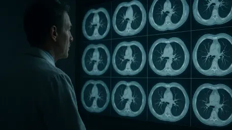
The technological core of COV-DSNet is rooted in a three-dimensional convolutional neural network, a significant departure from standard algorithms that frequently process medical images as a series of flat, disconnected slices. Standard two-dimensional systems often struggle to grasp the full extent of viral damage because pulmonary lesions are inherently three-dimensional structures that migrate and expand across various anatomical planes. By treating the entire chest computed tomography volume as a single spatial object, the 3D CNN effectively captures the volumetric density and distribution of ground-glass opacities and consolidations. This comprehensive view allows the system to identify the precise burden of the infection, which is often the most reliable indicator of whether a patient is heading toward recovery or collapse. Consequently, the AI provides a more holistic interpretation of lung pathology than even the most experienced radiologists could achieve through manual mental reconstruction.
Advanced Mechanisms: Mixed Convolution and Attention
To refine its diagnostic capabilities, the architecture incorporates mixed convolution and sophisticated attention mechanisms that mimic the focus of a trained specialist. Mixed convolution allows the network to strike a balance between broad spatial awareness and the extraction of fine-grained textural details within individual image slices, ensuring that small-scale damage is not overlooked during the analysis of the entire lung. Simultaneously, the attention mechanisms serve as computational filters that train the algorithm to prioritize diseased tissue while disregarding irrelevant anatomical structures like the heart, ribs, or major blood vessels. This dual approach minimizes imaging noise and helps the system pinpoint subtle inflammatory markers that might be obscured in traditional scans. By focusing only on the areas of the lung that contribute to the disease stage, the AI maintains a high level of accuracy even in complex cases where secondary health issues or imaging artifacts might otherwise confuse the diagnostic process.
Validating Accuracy and Clinical Reliability
Establishing the reliability of any new medical technology is a rigorous process that involves extensive testing against established clinical benchmarks. For COV-DSNet, this meant proving that an algorithm could accurately reflect the complex biological reality of a patient’s lungs as seen on a computed tomography scan. The validation phase was critical for demonstrating that the AI could distinguish between different phases of the illness with a high degree of certainty. In an environment where every medical decision can have profound consequences, the confidence of the clinical team in their diagnostic tools is paramount. The researchers focused on ensuring that the system’s outputs were not only accurate in a mathematical sense but also relevant to the practical needs of frontline physicians. By subjecting the model to a diverse range of patient data, the team aimed to create a robust tool that could perform consistently under the varied conditions of a real-world hospital setting.
Measuring Performance: Data and Predictive Power
The efficacy of this deep learning approach was rigorously tested using a substantial dataset of five hundred and seventy-eight patients, which was eventually narrowed down to a high-quality cohort of one hundred and fifty-six individuals. The research team focused on two hundred and ninety-seven specific scans to train the system in binary classification, distinguishing between the progression and remission stages of the disease. To measure the success of these predictions, the researchers utilized the area under the receiver operating characteristic curve, a standard statistical metric where higher values indicate superior diagnostic performance. COV-DSNet demonstrated a mean value of zero point eight two zero in its general analysis, which increased to zero point eight six four when applied to a dedicated verification set. These results confirm that the system is not only capable of processing large volumes of data but also provides a consistent and reliable staging tool that could be integrated into clinical workflows.
Clinical Value: The Importance of High Specificity
Perhaps the most significant clinical finding was the system’s high specificity, which reached a remarkable level of zero point nine two one during the verification phase. In the context of intensive care, high specificity is often more valuable than high sensitivity because it ensures that the system identifies patients in remission with exceptional accuracy. A diagnostic tool that frequently produces false alarms by predicting progression when a patient is actually improving could lead to the misallocation of resources, such as unnecessary ventilator use or the administration of toxic antiviral drugs. By correctly identifying when a patient’s lungs are beginning to heal, COV-DSNet empowers physicians to de-escalate care and transition patients toward recovery with a much higher degree of confidence. This ability to confirm remission objectively reduces the psychological and physical burden on patients while optimizing the use of critical hospital infrastructure during periods of high demand.
Future Implications and Prognostic Integration
The potential applications of deep learning in pulmonary medicine extend far beyond simple image classification, offering a glimpse into the future of integrated healthcare. As clinicians look for ways to improve patient outcomes, the ability to combine various types of medical data into a single, cohesive prognostic model has become increasingly valuable. COV-DSNet represents a foundational step in this direction by demonstrating how visual data from lung scans can be synthesized with physiological and laboratory markers. This multi-modal approach allows for a more comprehensive understanding of a patient’s overall health status and likely future trajectory. Furthermore, the insights gained from this specific application are helping to inform the development of similar tools for other respiratory conditions. By refining how we interpret the structural and functional changes in the lungs, we can prepare for a wide array of health challenges, ensuring that the medical community remains agile and informed in the face of emerging pathogens.
Beyond the Image: Enhancing Models and Viral Insights
One of the most powerful aspects of the study was the integration of AI staging with traditional clinical metrics like the APACHE II score and oxygenation ratios. When these data points were combined, the predictive accuracy for patient deterioration reached an unprecedented level of precision, suggesting that CT scans contain hidden visual biomarkers that blood tests alone cannot capture. This comprehensive modeling enabled clinicians to identify patients at the highest risk of failure well before traditional markers showed signs of decline. Additionally, the comparison between the AI’s structural analysis and RT-PCR viral loads revealed that lung damage does not always follow viral replication patterns. The system documented the ongoing inflammatory response of the body, providing a more accurate historical record of the disease’s impact than a standard viral swab. These insights proved essential for understanding the long-term recovery process and ensuring that treatment continued until structural healing was truly underway.
Addressing Limitations: A Roadmap for Universal Care
The researchers concluded that the implementation of COV-DSNet represented a significant advancement in the objective staging of viral pneumonia. They observed that by converting raw three-dimensional imaging data into actionable clinical stages, the system provided medical teams with the foresight needed to optimize resource allocation. Hospitals that adopted this technology reported a more efficient de-escalation of care for patients in the remission stage, which helped to alleviate the pressure on intensive care infrastructure. Furthermore, the study prompted a wider discussion on the necessity of external validation and the integration of AI as a standard decision-support tool. The clinical community recognized the value of these automated assessments in reducing the subjective variability of manual readings. Ultimately, the successful deployment of these deep learning models paved the way for more personalized treatment strategies and established a new benchmark for high-resolution diagnostic precision in the management of acute respiratory distress.