
Radiologists frequently observe breast arterial calcification during cancer screenings, yet fewer than two-thirds consistently document these findings in clinical reports. This oversight represents a significant missed opportunity in the realm of preventative medicine, particularly since cardiovascular disease remains the primary cause of mortality among women globally. While the focus of a mammogram is traditionally centered on the early detection of malignancy, the breast tissue serves as a window into the systemic health of the patient. The presence of breast arterial calcification, or BAC, is a potent biomarker for the buildup of plaque in other parts of the body, including the coronary arteries. Despite the clinical relevance of these findings, the sheer volume of cases and the lack of a standardized reporting framework often result in these vital clues being excluded from the final diagnostic report. As healthcare systems look toward 2026 and beyond, the integration of artificial intelligence is being viewed as the most viable solution to bridge this gap, ensuring that incidental findings are leveraged to improve long-term patient outcomes without adding to the administrative burden of already overextended radiology departments.
The Hidden Connection: Arterial Markers in Breast Tissue
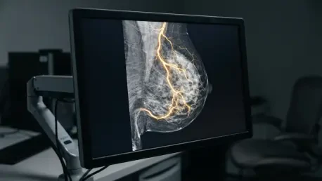
The biological significance of breast arterial calcification lies in its role as a visible indicator of medial arterial sclerosis, a condition where calcium deposits form within the arterial walls. Unlike the calcifications associated with breast cancer, which are typically found within the ducts or lobes, BAC is strictly vascular and does not indicate a risk of malignancy. However, its presence is far from benign when viewed through the lens of cardiovascular health. Modern clinical research has established a robust correlation between the extent of BAC and the risk of developing major adverse cardiovascular events, such as myocardial infarction or stroke. Because mammography is a routine part of healthcare for millions of women, it provides a unique, pre-existing data source that could be used to identify high-risk individuals long before they present with symptomatic heart disease. This opportunistic screening approach is particularly valuable because it requires no additional radiation exposure and no extra cost to the patient, utilizing images that have already been captured for oncological purposes.
The current challenge in the clinical environment is the inconsistency with which these findings are communicated to primary care physicians and cardiologists. Surveys indicate that while the vast majority of radiologists recognize the link between BAC and heart disease, only a small fraction consistently include this information in their reports. This disparity is often attributed to the high-pressure nature of modern diagnostic imaging, where the primary objective is to rule out cancer as quickly and accurately as possible. Without a mandate or an automated way to quantify these calcifications, many specialists feel that reporting BAC might lead to unnecessary patient anxiety or a flood of low-value referrals to cardiology. This is where specialized deep-learning modules come into play, offering a way to standardize the detection and reporting process. By automating the identification of vascular calcium, healthcare providers can ensure that every woman receives a comprehensive assessment of both her oncological and cardiovascular risk profiles during a single visit.
The Zurich Study: Evaluating Deep Learning in Practice
A landmark study conducted at the University Hospital of Zurich has recently provided a comprehensive evaluation of how artificial intelligence performs in a real-world screening environment. The research team utilized an experimental deep-learning module integrated into the b-box plus software package, analyzing an extensive dataset of 4,692 women and over 24,000 individual mammographic images. This retrospective analysis was designed to test the algorithm’s ability to identify BAC across a diverse population and to determine how various technical factors influenced its accuracy. The results highlighted a clear demographic trend: women who exhibited vascular calcifications were significantly older, with a median age of 68 compared to 56 for those without findings. This data reinforces the understanding that BAC is a progressive condition tied to aging and systemic vascular decline. By testing the AI against a manually reviewed subset of images, the study established a baseline for how automated tools can assist in identifying patients who may benefit from further cardiovascular evaluation.
The architecture of the AI system used in the Zurich study was trained on a global dataset, incorporating images from various hardware manufacturers and imaging techniques, including both 2D digital mammography and synthetic tomosynthesis. This diversity was critical for ensuring that the tool could remain effective across different clinical settings where equipment may vary. During the evaluation, the AI demonstrated a remarkably high specificity of 99 percent, meaning it was nearly flawless in its ability to correctly identify when calcifications were absent. While its sensitivity was more conservative at 35 percent, the high precision of the system ensures that it does not generate a high volume of false positives. In a clinical setting, this level of reliability is essential because it prevents the diagnostic workflow from being overwhelmed by inaccurate alerts. Instead, the AI acts as a high-confidence “rule-in” tool, flagging only the most certain cases of arterial calcification for further review by the medical team.
Performance Metrics: Precision versus Sensitivity
The discordance between human radiologists and AI algorithms often reveals the technical limitations and strengths of each. In the Zurich study, the majority of cases where the AI failed to detect calcifications involved very faint or low-intensity deposits that were only visible to the trained human eye. These false negatives were frequently associated with dense breast tissue, which can obscure vascular structures and make it difficult for an algorithm to distinguish between calcium and surrounding fibroglandular tissue. However, the rarity of false positives—only 8.5 percent of errors—suggests that the AI is highly effective at filtering out visual noise, such as surgical clips, implants, or skin lesions, which might otherwise be mistaken for arterial disease. Interestingly, there were instances where the AI identified legitimate calcifications that the human radiologist had initially overlooked, demonstrating the potential for these tools to serve as a reliable safety net in high-volume screening programs.
Beyond simple detection, the study delved into the “physics” of mammography to understand how image acquisition affects AI performance. Researchers discovered that the compression force applied during the procedure was a significant factor in the algorithm’s success. Higher compression levels thin the breast tissue and reduce overlying shadows, providing a clearer view of the arterial walls. This finding suggests that while AI is robust enough to handle moderate technical variability, optimized imaging protocols can further enhance its diagnostic power. The study also noted that standard quality metrics, such as the visibility of the pectoralis muscle or the nipple-to-pectoralis line, did not significantly impact the AI’s ability to see BAC. This level of robustness is encouraging for the widespread adoption of the technology, as it indicates that even slightly suboptimal images can still yield valuable cardiovascular insights without the need for a re-scan.
Clinical Implementation: From Research to Routine Care
The transition of AI from a research tool to a standard component of clinical practice requires a shift in how medical professionals view the mammography report. With the updated BI-RADS lexicon now officially recognizing the importance of documenting breast arterial health, the path is clear for a more holistic approach to women’s screening. Instead of being seen as a secondary or incidental finding, BAC is increasingly recognized as a vital sign for vascular wellness. Integrating AI into this workflow allows for the automatic generation of a cardiovascular risk score that can be sent directly to the patient’s primary care provider. This seamless data transfer ensures that the information is acted upon, leading to earlier interventions such as lipid-lowering therapies, lifestyle modifications, or more advanced cardiac imaging. By catching these signals early, healthcare systems can transition from a reactive model to a proactive one, potentially preventing thousands of cardiovascular events each year.
The strategic advantage of using an AI with high specificity is that it minimizes the potential for “over-diagnosis” and the subsequent medical costs associated with it. When the system flags a patient, the high positive predictive value gives clinicians the confidence they need to move forward with a referral. Furthermore, as these algorithms continue to evolve between 2026 and 2028, they will likely move beyond binary “yes/no” classifications to provide volumetric and density-based measurements of calcium. This would allow for a scoring system similar to the Agatston score used in cardiac CT scans, providing a more nuanced understanding of a patient’s risk level. The ability to track changes in arterial calcification over time, as a woman returns for her annual or biennial screening, would offer a longitudinal view of her cardiovascular health, making the mammogram one of the most versatile tools in the diagnostic arsenal.
Future Directions: Advancing Cardiovascular Risk Assessment
The medical community recognized that the integration of automated detection was no longer a luxury but a necessity for comprehensive preventative care. Experts determined that the most effective path forward involved the standardization of BAC reporting within the existing clinical frameworks to ensure consistency across different healthcare networks. Researchers suggested that future software iterations should focus on improving the sensitivity for detecting faint, early-stage calcifications while strictly maintaining the high specificity that prevented patient anxiety and unnecessary follow-up procedures. Healthcare administrators evaluated the long-term cost-effectiveness of these AI tools and found them to be a viable method for enhancing population health metrics without increasing the radiation dose or procedural complexity for the patient.
Clinicians moved toward a collaborative model where the mammography report served as a primary trigger for cardiovascular wellness checks. They established clear referral pathways that allowed women flagged by the AI to receive immediate consultations with specialists, effectively closing the gap between breast imaging and heart health. Scientists also explored the possibility of combining BAC data with other digital biomarkers, such as breast density and texture analysis, to create a multi-dimensional profile of a woman’s biological age. By the end of this developmental phase, the healthcare industry successfully transformed a routine cancer screening into a holistic diagnostic event, proving that the data already hidden within medical images held the key to significantly reducing the burden of heart disease in the female population.