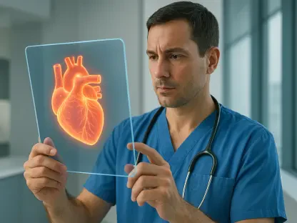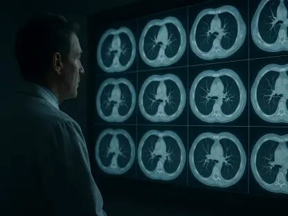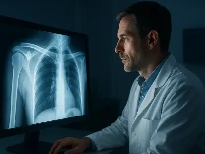

The development of xvr marks a shift toward unified surgical guidance where information from multiple imaging modalities is synthesized into actionable 3D data. Modern medicine has undergone a radical transformation through the rise of minimally invasive procedures, which allow surgeons to perform life-saving operations through incisions no larger than a dime. While these techniques significantly reduce patient recovery times and the risks of post-operative infections, they introduce a grueling cognitive challenge for the medical team. Surgeons must navigate the complex labyrinth of human vasculature or neurological pathways using two-dimensional X-ray imagery that lacks depth and spatial context. This forced clinicians to rely on years of specialized training to mentally reconstruct a three-dimensional environment from grainy, flat snapshots. The spatial gap between what the surgeon sees on the screen and the physical reality inside the patient often leads to increased procedural time and heightened stress during critical emergency interventions.
Overcoming Traditional Navigation Hurdles
The primary hurdle in modern catheter-based interventions is the accurate localization of surgical tools relative to delicate anatomical structures. When a clinician steers a thin wire through a patient’s heart or brain, they must know its position with absolute certainty to avoid catastrophic complications. Historically, this has required a process known as manual registration, where the surgical team attempts to align live 2D X-ray feeds with 3D medical scans, such as CT or MRI, that were obtained prior to the operation. This manual approach is notoriously slow and subjective, often forcing surgeons to pause during a procedure to input coordinates or identify landmarks on a digital display. In high-pressure environments like emergency stroke units, these pauses are more than just inconveniences; they represent lost time that can lead to permanent tissue damage. The lack of a seamless, automated bridge between 2D visuals and 3D maps has remained a persistent bottleneck in surgical suites.
Localization: The Critical Need for Spatial Accuracy
A significant limitation of current image-guided surgery is the “flatness” of the intraoperative perspective. While pre-operative CT scans provide a rich, three-dimensional view of the patient’s internal anatomy, the live guidance used during the actual operation is almost exclusively two-dimensional. This discrepancy creates a massive cognitive load, as the surgeon must constantly translate the two-dimensional movement of their instruments into a three-dimensional space they cannot actually see in real-time. Even with advanced training, the risk of spatial disorientation remains, particularly when navigating through tortuous vessels or near critical nerve clusters. The introduction of xvr seeks to eliminate this guesswork by providing a continuous, high-fidelity 3D map that updates as the surgeon moves. By creating a digital bridge between the pre-operative planning phase and the live surgical execution, the system ensures that the most detailed anatomical information is always available exactly when it is needed most.
Robustness: Challenges with Anatomical Diversity
Existing artificial intelligence models designed for image registration have frequently struggled with the inherent variability of human anatomy. Every patient presents a unique internal architecture, influenced by age, genetics, and specific pathologies that can distort standard biological landmarks. A model trained on a generalized group of subjects often fails when confronted with an individual whose anatomy falls outside the narrow parameters of the training data. This lack of robustness has prevented many automated tools from moving beyond the laboratory and into the operating room. Surgeons require a system that is not only fast but also incredibly reliable across a diverse demographic spectrum, including pediatric patients and those with rare structural anomalies. Without this consistency, the adoption of AI-assisted navigation remained limited to specialized research institutions rather than becoming a standard of care. The new system addresses this by using patient-specific data to ensure the model matches the person on the table.
The Innovation of Patient-Specific Modeling
The innovation of the xvr system stems from its departure from a universal AI architecture toward a patient-specific modeling framework. Instead of asking the AI to understand every human body simultaneously, the system creates a digital twin specifically tailored to the individual on the operating table. This workflow begins by taking the patient’s preoperative 3D scan and running it through a physics-based simulation. This simulation generates thousands of synthetic X-rays from virtually every conceivable angle at a rate of approximately 1,000 images per second. By anchoring the data generation process in the actual physics of X-ray transmission and the patient’s unique anatomical data, the researchers effectively eliminated the risk of hallucinations. These hallucinations, where AI creates visual artifacts that do not exist in reality, have been a major safety concern in medical imaging, but the xvr system ensures that every 3D projection is grounded in physical truth, making it a reliable clinical tool.
Simulation: Physics-Based Data and Accurate Projections
By grounding the synthetic data in the actual physics of radiation and human tissue density, the system creates a training set that is perfectly aligned with the patient’s real-world anatomy. This level of customization allows the AI to learn the specific nuances of a patient’s skeletal structure or vascular network before the surgery even begins. The ability to generate 1,000 realistic images per second means that the system can cover a vast range of possible viewing angles, ensuring that no matter how the X-ray C-arm is positioned during surgery, the AI has a corresponding 3D reference ready. This proactive data generation removes the need for the AI to “guess” the depth of an object based on generic human averages. Instead, the system recognizes the specific shadow and density patterns unique to that individual. This transition from general probability to specific physical simulation represents a major technological milestone in the reliability of medical machine learning models for surgical use.
Adaptation: Rapid Processing for Emergency Interventions
To make this patient-specific approach viable for emergency scenarios, the development team integrated a sophisticated foundation model capable of rapid adaptation. Pretrained on a massive dataset of whole-body 3D scans from more than 2,000 diverse patients, the foundation model possesses a deep understanding of general human structure. When a new patient arrives in the emergency room, the system does not need to start its learning process from the beginning. Instead, it uses the foundation model as a baseline and adapts it to the current patient’s specific anatomy in roughly five minutes. This represents a significant leap from previous iterations of the technology, which often required twelve hours or more for similar training cycles. Once this brief adaptation period is complete, the model can match live 2D X-rays to the 3D volume in a matter of seconds, providing the immediate feedback loop necessary for time-sensitive procedures like opening a blocked artery or treating a life-threatening stroke.
Impact on Clinical Performance and Accessibility
Rigorous validation of the xvr system involved testing it against the largest available dataset of real 2D-to-3D registrations, sourced from five different medical institutions. The study covered a wide array of anatomical structures, including complex bone groups and various organ systems in both adult and pediatric populations. The results were definitive, showing that xvr outperformed existing AI-driven methods by an order of magnitude. Most importantly, the system achieved sub-millimeter precision, which is the gold standard for navigating the tiny, fragile vessels found in the brain or neonatal cardiac systems. This level of accuracy remained consistent regardless of the patient’s age or specific medical condition, proving that the patient-specific adaptation method successfully navigates the challenge of anatomical diversity. The reliability of these results across different hospitals suggests that the technology is ready for large-scale clinical deployment in various surgical and interventional settings.
Results: Establishing Sub-Millimeter Precision Standards
The empirical evidence gathered during the testing phase highlighted the system’s ability to maintain high precision even in suboptimal imaging conditions. In many surgical scenarios, X-ray images can be obscured by surgical tools, medical implants, or low contrast, which traditionally confuses automated registration systems. However, because xvr was trained on the patient’s specific 3D volume, it remained highly resilient to these visual disruptions. The achievement of sub-millimeter accuracy is particularly vital for neurological interventions, where a deviation of even a single millimeter can result in damage to critical brain tissue. By proving that the AI could meet this stringent requirement across a broad range of real-world clinical data, the researchers demonstrated that the system is not just a theoretical improvement but a practical solution for the most demanding medical tasks. This level of performance established a new benchmark for what is possible in the field of automated image registration and surgical navigation.
Outlook: Advancing Toward Dynamic Surgical Robotics
The successful implementation of xvr demonstrated how advanced machine learning could bridge the gap between complex diagnostic imaging and real-time surgical action. By prioritizing patient-specific data over generic training models, the research team offered a viable solution to the long-standing problem of anatomical variability in medical AI. Clinicians who utilized these tools observed a marked improvement in their ability to navigate internal pathways without the constant need for manual recalibration. This advancement prioritized the safety of the patient while simultaneously reducing the cognitive load on the surgical staff during high-stakes operations. The project emphasized the importance of grounding generative models in physical laws to ensure clinical reliability. Hospitals began to evaluate how these rapid adaptation models could be integrated into existing emergency protocols to provide expert-level navigation in smaller, regional facilities that lacked specialized surgical centers.