
For decades, the invisible barrier of biological imaging has forced a brutal compromise between observing a cell’s life or preserving its structural integrity. The “water-window” spectral range, spanning 2.3 to 4.4 nanometers, provides a unique solution to this dilemma by utilizing a specific physical phenomenon. Within this narrow band, water is essentially transparent, while carbon—the primary building block of organic life—is opaque. This allows researchers to capture high-contrast images of living specimens in their natural, hydrated state without the destructive preparation required by other high-resolution methods.
Historically, capturing these specific wavelengths required access to synchrotrons, which are massive particle accelerators the size of sports stadiums costing billions of dollars. The transition toward laboratory-scale solutions is a transformative shift that brings the power of centralized facilities directly into individual research centers. By miniaturizing the technology, the industry is moving away from exclusive, rare-access sites to a decentralized model that prioritizes immediate, real-time observation. This democratization of high-contrast imaging is vital for the next decade of biological discovery.
Core Technical Components and Innovations
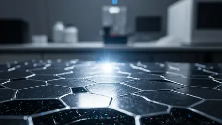
Graphite Nanoantenna Arrays
The technical foundation of modern tabletop water-window generation relies on ultrathin graphite flakes, typically ranging from 10 to 170 nanometers in thickness. Graphite’s crystalline atomic structure is not merely a material choice but a functional component that acts as a coordinated array of antennas. When low-energy electrons strike these flakes, the periodic arrangement of carbon atoms directs the resulting electromagnetic radiation with extreme precision.
This implementation is unique because it leverages the natural geometry of the carbon lattice to synchronize the emission of X-rays. Unlike traditional targets that produce scattered, incoherent radiation, the graphite nanoantenna array ensures that the output is concentrated and directed. This coordination allows for higher efficiency in photon production, which is essential for maintaining image quality in a much smaller physical footprint than previously possible.
Low-Energy Electron Emission Systems
The replacement of massive particle acceleration tubes with low-energy electron emission systems has fundamentally altered the economics of X-ray generation. Traditional synchrotrons require high-velocity particles to generate the necessary energy, which necessitates hundreds of meters of shielding and vacuum tubes. In contrast, the new tabletop systems use specialized cathodes to emit low-energy electrons that interact with the graphite target to produce the same spectral results.
This miniaturization significantly reduces the physical footprint and the energy consumption of the imaging process. By optimizing the electron-to-photon conversion efficiency, these systems allow for high-intensity output without the heat management issues that plagued earlier attempts at lab-scale X-rays. This makes the technology viable for standard research laboratories that lack the infrastructure for heavy industrial machinery.
Precise Wavelength Tunability
One of the most significant advantages of this new generation of X-ray sources is the ability to adjust the wavelength with high precision. By modulating the speed of the incoming electrons and the orientation of the graphite flakes, researchers can fine-tune the X-ray energy. This tunability is critical for distinguishing between various biomolecules, such as proteins and lipids, which have slightly different absorption signatures within the water window.
Compared to traditional fixed-energy laboratory machines, which offer a “one-size-fits-all” approach, tunable sources provide a surgical level of detail. This flexibility allows for the identification of specific cellular components during dynamic processes. The ability to shift wavelengths on demand means that a single machine can serve multiple research functions, from basic pathology to advanced material analysis, without hardware reconfigurations.
Recent Advancements in Tabletop Generation
Recent breakthroughs, particularly those led by institutions like Nanyang Technological University, have solved the stability issues that once hindered tabletop adoption. Previous iterations often relied on high-intensity lasers that were prone to fluctuations and required constant maintenance. The shift to a laser-free, electron-driven approach has created a more stable and reliable imaging environment. This stability ensures that long-exposure imaging can be conducted without the risk of data loss or beam degradation.
Moreover, innovations in filtering technology have allowed for the isolation of water-window radiation with unprecedented purity. By removing unwanted higher-energy “noise,” the new prototypes achieve a signal-to-noise ratio that rivals synchrotron data. The transition from proof-of-concept experiments within electron microscopes to standalone, portable prototypes signifies that the technology is moving out of the purely academic realm and into practical industrial production.
Real-World Applications and Sector Integration
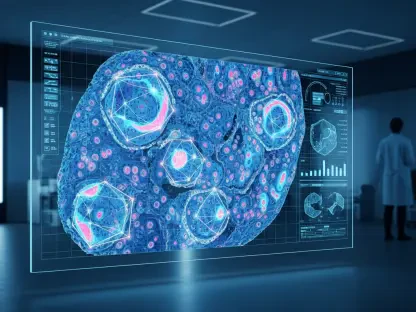
Real-Time Biological Research and Pathology
The primary impact of water-window technology is the ability to observe dynamic cellular processes as they occur. Scientists can now watch a virus invade a host cell or track the movement of organelles in a living environment. Because the process eliminates the need for dehydration or chemical staining—both of which kill the sample and alter its structure—the resulting data is a more accurate representation of biological reality.
This advancement is particularly crucial for studying disease progression and evaluating the efficacy of new drug treatments. By observing how a therapeutic intervention affects a live cell in real-time, researchers can identify potential side effects or mechanical failures much earlier in the development cycle. This shift toward live, high-resolution pathology is expected to accelerate the pace of medical breakthroughs significantly.
Material Science and Sustainable Energy
Beyond biology, the technology is revolutionizing the inspection of advanced semiconductors and microelectronics. As components shrink to the nanometer scale, traditional X-rays lack the contrast to identify defects in light-element structures like photoresists or organic polymers. Water-window X-rays provide the resolution needed to inspect internal layers of chips without damaging the delicate circuits.
In the sector of sustainable energy, this technology is being used to optimize the next generation of battery structures and solar cell layers. High-resolution imaging allows engineers to see how lithium ions move through an anode or how light is absorbed within a thin-film solar panel. These insights lead to material optimizations that are essential for the global transition to green energy, making X-ray technology a key driver of industrial sustainability.
Clinical Diagnostics and 3D Biopsies
The potential for high-resolution, three-dimensional biopsies in hospital settings represents a major leap in precision medicine. Current clinical X-rays provide a flat, low-contrast view of soft tissues, often requiring painful invasive procedures to confirm a diagnosis. Water-window imaging could allow for non-destructive 3D visualization of tissue samples at a molecular level, providing doctors with a “virtual slice” of a tumor or organ.
This technology offers a level of detail that standard medical X-rays simply cannot match, especially regarding soft tissue contrast. While it is not a replacement for full-body radiography, it serves as a specialized tool for pathology labs and surgical suites. The integration of 3D molecular imaging into the clinical workflow will likely redefine how doctors approach complex diagnoses in the coming years.
Current Challenges and Adoption Barriers
Despite its promise, the technology faces technical hurdles regarding the scaling of prototypes for mass commercial production. Engineering a device that maintains synchrotron-level precision while being operated by non-specialized staff in a hospital or industrial plant requires significant refinement. Ensuring the longevity of the graphite targets under continuous electron bombardment remains a primary focus for current research teams.
Navigating the complex regulatory landscape for medical devices is another significant barrier to immediate adoption. Clinical approval processes are rigorous and time-consuming, meaning the transition from the laboratory to the patient bedside will be a multi-year journey. Additionally, while the technology is cheaper than a synchrotron, the initial investment costs for small research institutions remain high, requiring innovative financing models to ensure widespread access.
Future Outlook and Technological Trajectory
The trajectory of water-window technology points toward the total democratization of high-resolution imaging. As manufacturing costs decrease, the availability of these devices will likely shift the paradigm of molecular research from a few centralized hubs to a global network of local laboratories. This change will allow for faster response times during public health crises and more localized innovation in materials science.
In the long term, we can expect a shift in how laboratories and hospitals manage molecular-level research. The integration of artificial intelligence with real-time X-ray data will likely lead to automated molecular discovery, where machines can identify new viral structures or material defects instantly. This evolution will further reduce the carbon footprint of particle-based imaging, aligning high-level science with global sustainability goals.
Summary and Assessment of Technological Impact
The transition from billion-dollar facilities to accessible tabletop devices marked a fundamental shift in imaging capabilities. This evolution moved high-resolution molecular research from exclusive, centralized hubs into the hands of individual laboratories. The technology successfully bridged the gap between electron microscopy and standard radiography by providing high-contrast results in hydrated environments without damaging the specimens.
The readiness for industrial and clinical integration proved to be the most significant milestone for this sector. While challenges in mass production remained, the overall significance of water-window X-rays became undeniable as a tool for both medicine and material science. This advancement provided a smaller, more affordable, and sustainable way to see the fundamental building blocks of the world, effectively closing a long-standing gap in scientific observation.