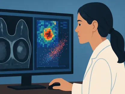

Deep learning systems are overcoming the technical challenges of fetal MRI by processing images regardless of the orientation or specific characteristics of the scan. This technological leap represents a significant turning point in prenatal medicine, offering a way to bypass the grueling manual tasks that have long slowed down critical diagnoses. Researchers at Cincinnati Children’s Hospital Medical Center recently unveiled a study in the journal Pediatric Radiology that details the creation of an automated segmentation tool designed to evaluate fetal lung health with unprecedented precision. By utilizing vast datasets and sophisticated neural architectures, this system addresses a primary bottleneck in fetal imaging: the quantification of lung volumes in high-risk pregnancies. As medical centers worldwide strive to provide more accurate prognostic information to expectant parents, the integration of artificial intelligence into the radiology suite emerges as a vital strategy for improving both clinical workflow and patient outcomes during the most delicate stages of life.
The Clinical Urgency: Understanding Pulmonary Hypoplasia
The Challenge: Quantifying Life-Saving Metrics
Quantifying fetal lung development serves as the primary method for predicting pulmonary hypoplasia, a condition where the lungs are insufficiently developed to maintain life after delivery. This life-threatening state often arises as a final common pathway for several serious fetal anomalies, most notably Congenital Diaphragmatic Hernia. In these cases, a defect in the diaphragm allows abdominal organs to migrate into the thoracic cavity, physically compressing the lungs and hindering their growth. Beyond hernia cases, pulmonary hypoplasia can also result from renal malformations that cause low amniotic fluid levels, bladder outlet obstructions, or restrictive skeletal dysplasias that prevent the chest cavity from expanding properly. To manage these high-risk scenarios, medical teams rely on Total Lung Volume measurements derived from MRI scans to determine whether a fetus requires specialized surgical interventions or if the family should prepare for a delivery at a tertiary care center equipped with advanced life support systems.
The Bottleneck: Manual Labor in High-Stakes Environments
Despite the critical nature of these measurements, the traditional method of calculating lung volume is notoriously labor-intensive and prone to human error. Radiologists must manually trace the boundaries of the lungs on every individual slice of an MRI scan, a process that can take thirty minutes or even an hour for a single patient. This timeline is often extended when the fetus is particularly active or when the anatomy is severely distorted by herniated organs, making it difficult to distinguish between lung tissue and other structures. Because of the immense time commitment required, even the most advanced imaging centers are limited in the number of cases they can process each day. Furthermore, the subjective nature of manual tracing means that two different experts might arrive at slightly different volume estimates, introducing a level of variability that can complicate sensitive clinical decisions and parent counseling sessions during a time of extreme emotional stress.
Technical Breakthroughs: The Architecture of Intelligence
Robust DatTraining for Real-World Complexity
To overcome the limitations of previous automation attempts, the research team utilized a massive retrospective dataset of over 1,200 fetal MRI examinations conducted over the last decade leading up to 2026. They meticulously selected 906 high-quality examinations that had been used in real-world clinical counseling to ensure the AI was trained on diverse and representative data. This collection spanned gestational ages from 16 to 38 weeks and included a wide spectrum of pathologies, moving far beyond the “normal” lung scans that limited earlier models. By training the AI on such a varied dataset, the researchers ensured that the system could handle the complex anatomical presentations found in patients with renal disease, skeletal dysplasia, and varying degrees of diaphragmatic defects. This inclusive approach to data selection is what allows the model to remain reliable across different phenotypes, making it a truly versatile tool for a modern, high-volume prenatal care facility.
Performance Metrics: Efficiency Meets Accuracy
The core of this innovation lies in the nnU-Net v2 framework, a state-of-the-art deep learning architecture that handles the intricate preprocessing required for medical imaging. Using high-performance NVIDIA A100 GPUs, the researchers developed an ensemble model that processes standard coronal T2-weighted images almost instantaneously. In testing, the AI completed the segmentation of Total Lung Volume in an average of 3.8 seconds, representing a 450-fold increase in efficiency compared to manual methods. Statistically, the system achieved an Intraclass Correlation Coefficient of 0.955, a score that indicates “excellent” agreement with human experts. With a median absolute percentage error of only 11.1%, the AI’s performance mirrors the natural variability seen between two experienced radiologists. This level of speed and precision allows clinicians to obtain results while the patient is still at the imaging facility, fundamentally changing how quickly a medical team can pivot toward a treatment plan.
Clinical Implementation: Moving from Laboratory to Clinic
The Collaborative Workflow: Human-in-the-Loop Paradigm
Rather than seeking to replace the expertise of medical professionals, the research team designed the system to function within a “human-in-the-loop” workflow. In this model, the AI performs the heavy lifting by generating the initial lung tracings in seconds, which the radiologist then reviews and refines if necessary. Simulations showed that even in the most difficult cases, a physician could correct the AI’s output in just over three minutes, still resulting in a 90% reduction in total labor time. This collaborative approach ensures that the final diagnostic volume remains under the oversight of a trained expert while removing the repetitive, fatigue-inducing parts of the task. By streamlining the process so significantly, hospitals can handle a much higher volume of complex cases without increasing the burden on their staff. This efficiency is particularly valuable for multidisciplinary teams who must coordinate quickly to plan for high-risk deliveries and immediate neonatal surgeries.
Pathological Stability: Handling Complex Phenotypes
One of the most impressive aspects of the new AI system is its stability across different disease types and suboptimal scanning conditions. The model remained highly accurate even when faced with skeletal dysplasias that restrict the thoracic cage or renal conditions that result in minimal amniotic fluid, which often degrades image contrast. While the system faced some challenges in distinguishing the thymus gland from hypoplastic lung tissue in very early pregnancies or in cases with massive lung lesions, its performance was overall remarkably consistent. The study highlighted that the AI is particularly effective during the critical clinical windows when surgical decisions are typically made. This reliability across various phenotypes ensures that the technology can be deployed in a wide range of clinical scenarios, providing a standardized baseline for measurement that is not affected by the specific orientation of the fetus or the technical nuances of the MRI sequence being used.
Strategic Outlook: Future Directions in Prenatal Care
Global Access: Standardizing Quantitative Assessment
The successful validation of this deep learning system at a major medical center paved the way for a global shift toward standardized prenatal diagnostics. By automating the most difficult aspect of fetal lung assessment, the technology allows smaller regional hospitals to provide the same level of quantitative precision as elite tertiary centers. This democratization of data-driven medicine ensures that no matter where a patient is located, they can receive a high-quality prognostic evaluation based on objective metrics rather than local expertise alone. Moving forward, the focus shifted toward multi-institutional validation, ensuring the AI performs consistently across different scanner brands and software versions. As these systems become more integrated into standard hospital software, the once-specialized task of lung volumetry became a routine part of the prenatal imaging protocol, allowing doctors to focus more on patient care and less on the technical hurdles of image processing.
Actionable Progress: Advancing the Field of Radiomics
The development of this rapid segmentation tool also serves as a foundational step for the expanding field of fetal radiomics, where researchers analyze large-scale imaging data to find hidden patterns in disease progression. Because the AI provides highly reproducible results, it enables the creation of massive, standardized databases that were previously impossible to assemble through manual labor. These datasets allow scientists to refine growth charts and improve the predictive power of lung volume measurements for a variety of rare conditions. In the years following this research, clinical teams transitioned toward these automated solutions to facilitate same-day results, drastically reducing the psychological burden on families waiting for answers. The integration of such tools into the daily workflow allowed for more frequent monitoring of high-risk pregnancies, ensuring that any changes in fetal development were identified and addressed with immediate, life-saving interventions as soon as the data became available.