
The historical reliance on static, destructive snapshots for evaluating cancer therapies has finally been superseded by a sophisticated UCLA-led platform that merges high-throughput 3D bioprinting with label-free imaging and artificial intelligence. This shift represents a significant departure from traditional methodologies that often require the destruction of biological samples to extract meaningful data. By facilitating the creation of patient-derived tumor organoids within a controlled environment, this technology allows for the continuous observation of cellular behavior over extended periods. Researchers can now monitor the dynamic evolution of a tumor’s response to various pharmaceutical interventions without the interference of chemical labels or dyes. This comprehensive approach addresses the limitations of conventional screening, offering a more nuanced understanding of how cancer cells interact with their environment and survive treatment. The integration of these technologies creates a high-speed workflow that prioritizes biological integrity.
Automating Consistency: The Integration of Bioprinting in Oncology
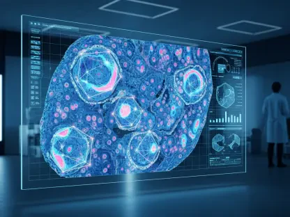
The automation of tumor organoid production marks a pivotal advancement in the quest for laboratory consistency and high-throughput screening capabilities. Manual cultivation of organoids has long been fraught with human error and variability, leading to inconsistent results that hinder the development of reliable cancer therapies. By utilizing specialized 3D bioprinting systems, scientists have successfully automated the fabrication of thousands of identical tumor models simultaneously. This process involves the precise deposition of cells and extracellular matrix materials into organized structures that mimic the three-dimensional architecture of actual tumors. The ability to produce such high volumes of uniform samples ensures that any observed differences in drug response are due to the treatment itself rather than variations in the organoid’s construction. Consequently, this level of standardization accelerates the pace of research by allowing for more ambitious experimental designs.
Standardized production through bioprinting also facilitates the exploration of complex tumor microenvironments that were previously difficult to replicate in a laboratory setting. By precisely controlling the spatial arrangement of various cell types, including immune cells and fibroblasts, researchers can create more realistic models of human cancer. This structural fidelity is crucial for understanding how the surrounding tissue influences a tumor’s growth and its susceptibility to medication. Furthermore, the high-throughput nature of this automated system allows for the simultaneous testing of vast chemical libraries against identical tumor structures. This capability significantly reduces the time and resources required to identify promising new drug candidates, as thousands of variations can be analyzed in parallel. The resulting data provides a robust foundation for subsequent clinical trials, ensuring that only the most effective treatments move forward. This move enhances preclinical data reliability.
Advancing Observation: The Power of Label-Free Quantitative Phase Imaging
Quantitative phase imaging stands as a cornerstone of this new platform, offering a non-invasive alternative to the destructive staining methods traditionally used in cellular biology. This technique leverages the physical properties of light as it passes through biological specimens, measuring the minute shifts in phase that occur when photons interact with cellular components. Because this method does not require the addition of fluorescent dyes or radioactive labels, the biological integrity of the tumor organoids remains uncompromised throughout the imaging process. This preservation of cell health is vital for long-term studies, as it allows for the continuous monitoring of the same live samples over many days. Traditional methods often terminate a sample at a single time point to measure its biochemical state, providing only a limited perspective on a treatment’s efficacy. In contrast, label-free imaging provides a constant stream of data, capturing the entire trajectory of the tumor’s reaction.
The longitudinal nature of label-free imaging enables researchers to observe transient cellular behaviors that are often missed by conventional endpoint assays. For instance, the initial response of a tumor to a specific therapy can differ significantly from its long-term survival strategy, and seeing this progression in real-time is essential for identifying drug resistance. By tracking the morphological changes and mass distribution within an organoid, scientists can pinpoint exactly when and where a treatment begins to fail. This high-resolution temporal data allows for a more detailed analysis of the cell cycle and the metabolic shifts that occur during drug exposure. Moreover, because the imaging process is entirely non-destructive, the organoids can be reused for further analysis or even sequential drug testing. This efficiency not only saves biological material but also provides a more comprehensive narrative of how cancer cells adapt to changing environmental pressures throughout the study.
Intelligent Analytics: Deciphering Complex Drug Responses through Deep Learning
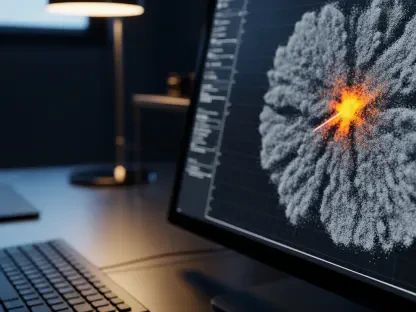
The sheer volume of visual data generated by continuous label-free imaging necessitates the use of advanced artificial intelligence to extract meaningful biological insights. Deep learning algorithms are uniquely equipped to process these massive datasets, identifying subtle patterns in cell growth and movement that would be imperceptible to the human eye. These AI models are trained on thousands of images to recognize the distinct signatures of healthy versus dying cells, allowing for the automated quantification of drug efficacy. By removing the subjectivity often associated with manual image analysis, machine learning ensures that the resulting data is both objective and reproducible across different research facilities. This computational approach also allows for the simultaneous tracking of multiple parameters, such as changes in organoid volume, density, and overall structural complexity. As the algorithms learn from new data, their predictive accuracy improves, making them indispensable for research.
The medical community successfully established a standard framework for the integration of bioprinted organoids into clinical pathology workflows. Stakeholders prioritized the development of interoperable AI platforms that allowed disparate laboratory systems to communicate and share longitudinal data effectively. Researchers implemented rigorous validation protocols that ensured bioprinted models accurately reflected the metabolic and structural characteristics of original patient tumors. These steps were essential in moving beyond experimental prototypes into reliable diagnostic tools that supported real-time therapeutic adjustments. Furthermore, academic institutions expanded their curricula to include cross-disciplinary training in bioengineering and machine learning, ensuring a steady pipeline of experts. The scientific body also adopted ethical guidelines for the storage and use of patient-derived samples. These efforts secured the role of bioprinting as a permanent pillar of oncology for the coming years.