
The oncology landscape has reached a critical juncture where the volume of imaging data generated during cancer treatment frequently outpaces the human capacity to interpret it with the required precision. Clinical radiologists often find themselves navigating a sea of complex scans, where measuring tiny changes in tumor volume or identifying subtle shifts in metastatic spread can mean the difference between a successful intervention and a missed opportunity. To address these systemic inefficiencies, Raidium has introduced a specialized platform built from the ground up to leverage artificial intelligence as a core component rather than a mere add-on to existing legacy systems. By integrating deep learning models directly into the diagnostic workflow, this technology aims to provide clinicians with highly accurate, automated longitudinal tracking that allows for a more nuanced understanding of disease progression. This shift toward AI-native environments represents a fundamental change in how medical data is synthesized.
Evolution of Diagnostic Accuracy: Enhancing Clinical Precision
Automated Volumetric Assessment: Beyond Linear Metrics
Traditional methods of measuring tumors rely heavily on manual two-dimensional cross-sections, a process that is not only time-consuming but also prone to significant inter-observer variability between different clinicians. The introduction of Raidium’s platform changes this dynamic by employing sophisticated computer vision algorithms that can automatically segment and measure the entire volume of a lesion in three dimensions. This level of automation ensures that measurements are consistent regardless of which radiologist is reviewing the scan, providing a more reliable foundation for assessing therapeutic efficacy over time. When a patient undergoes multiple rounds of chemotherapy or radiation, tracking the volumetric change in a tumor provides a far more accurate reflection of the biological reality than simple linear diameters. Furthermore, the system is designed to detect the emergence of new nodules that might be overlooked during a standard review, effectively acting as a high-fidelity safety net for medical teams.
Workflow Integration: Reducing the Cognitive Burden
Efficiency in the radiology suite is further enhanced by the platform’s ability to pre-process images before a physician even opens the study, effectively staging the data for immediate clinical review. Instead of spending valuable minutes performing routine segmentations and cross-referencing previous studies, the radiologist is presented with a comprehensive summary of changes that have occurred since the last visit. This proactive approach reduces the cognitive load on healthcare providers, allowing them to dedicate more time to the complex interpretative tasks that require human judgment and specialized expertise. By standardizing the way oncological data is presented and analyzed, the platform facilitates clearer communication between radiologists and referring oncologists. This collaborative transparency is essential in high-stakes environments where treatment adjustments must be made rapidly based on imaging findings. The system also minimizes the likelihood of clerical errors as the automated metrics are directly populated into records.
Systemic Interoperability: Future-Proofing Medical Infrastructure
Cross-Modal Data Harmonization: A Unified Clinical View
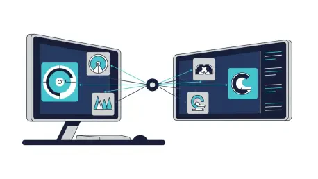
Modern oncology relies on a diverse array of imaging modalities, ranging from traditional computed tomography to advanced magnetic resonance imaging and positron emission tomography scans. Raidium’s infrastructure is engineered to ingest these disparate data streams into a unified environment, where cross-modal comparisons can be conducted with unprecedented ease and technical accuracy. The platform utilizes advanced registration techniques to align scans taken at different times or via different machines, ensuring that the spatial orientation of a tumor is perfectly matched for longitudinal analysis. This capability is particularly vital when dealing with complex metastatic cases where lesions may be scattered across different organ systems and require varying imaging protocols for optimal visualization. By consolidating these views, the platform provides a holistic perspective on the patient’s condition that was previously difficult to achieve without manual intervention. This technical cohesion allows for a more comprehensive assessment of responses.
Strategic Implementation: Practical Steps for Clinical Teams
The move toward AI-native radiology platforms marked a significant departure from the siloed and manual workflows that had characterized oncology for several decades. As the volume of medical imaging continued to expand, the necessity for tools that could synthesize information at scale became undeniable for maintaining high standards of patient care. Organizations that adopted these advanced platforms positioned themselves at the forefront of a major technological shift, moving away from reactive diagnostics toward a more predictive and data-driven model of treatment. This transition not only improved the accuracy of tumor tracking but also provided a wealth of structured data that was used for retrospective research and the development of new clinical protocols. Healthcare providers eventually leveraged these insights to create more personalized care plans, ensuring that every patient received a treatment strategy tailored to their specific biological response. The focus shifted from mere observation to active, data-informed intervention strategies.