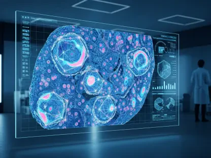

The traditional medical approach to breast cancer screening has long functioned as a snapshot in time, capturing only the immediate visible state of the patient’s health rather than the complex biological narrative unfolding beneath the surface. Recent advancements in oncology are fundamentally shifting how clinicians evaluate long-term risk, moving away from static assessments toward dynamic, data-driven predictions. A landmark study published in the journal Radiology illustrates that tracking artificial intelligence scores over several years can predict future cancer with significantly higher precision than traditional methods. This innovative approach allows the medical field to transition toward a more personalized strategy, focusing on catching subtle warning signs before they ever become apparent to even the most experienced human eye. By leveraging deep learning, doctors can now identify micro-patterns in tissue that suggest a trajectory toward disease, providing a crucial head start in a race where time is the most valuable resource for survival.
Analyzing the DatHow AI Tracks Patient Risk
Breakthroughs: Longitudinal Screening and Data Analysis
The foundation of this technological shift rests on a massive longitudinal dataset consisting of more than 54,000 women and nearly 240,000 mammograms collected over the span of a decade. Researchers discovered that there is a profound and measurable difference between the imaging data of women who eventually developed cancer and those who remained healthy. While a single mammogram might appear normal to a radiologist, the AI algorithms were able to detect microscopic signatures that suggested the presence of developing ductal carcinoma in situ or invasive cancer years before a physical tumor formed. This research proved that the risk of developing breast cancer is not a random event but often a slow-moving biological process that leaves a trail of digital evidence. By analyzing multiple scans over time, the AI identifies whether a patient’s breast tissue is maintaining a stable health profile or if it is beginning to exhibit the early characteristics of malignancy.
Building on these findings, the study demonstrated that AI-generated risk scores provide a much more reliable metric than traditional factors like family history or current breast density alone. In the past, these factors were treated as fixed variables, but the AI treats every new mammogram as an update to a living health record. For the women in the study who eventually received a diagnosis, their AI risk scores showed a steady and measurable climb over a six-year period. In contrast, the group that remained cancer-free exhibited scores that were consistently low and stable throughout the entire ten-year observation window. This clear divergence in data points allows healthcare providers to categorize patients not just by their current state, but by their “risk trajectory.” This capability represents a monumental leap in diagnostic science, as it replaces generalized population statistics with a highly specific, individualized analysis of how a particular patient’s breast tissue is evolving.
The Temporal Gradient: Identifying Early Warning Signs
One of the most striking revelations from the research was the identification of what scientists call a “preclinical window,” a period where the AI spots changes that are otherwise invisible. The data indicated that while the rise in risk scores was gradual over several years, it accelerated sharply in the twenty-four months leading up to a clinical diagnosis. This acceleration serves as a vital red flag, signaling that the biological environment is shifting rapidly toward a state that supports tumor growth. Identifying this temporal gradient gives clinicians a specific window for intervention that simply did not exist under previous screening protocols. By recognizing these sharp upward trends in the AI score, doctors can move a patient from standard annual observation to a high-priority monitoring status. This proactive stance ensures that if a tumor does manifest, it is caught at its earliest, most treatable stage, or potentially prevented through prophylactic measures tailored to the patient’s profile.
Furthermore, this ability to monitor the rate of change in tissue health provides a solution for identifying “sporadic” breast cancers, which are cases that occur in women without any known genetic predispositions. Historically, these patients often fell through the cracks of risk-assessment models because they lacked the traditional markers for high risk, such as the BRCA gene mutation. The AI’s focus on the actual biological behavior of the tissue rather than purely on hereditary factors allows for a much broader net to be cast. This level of insight is particularly effective because it bypasses the limitations of human visual perception, which is often hindered by the subtle nature of early-stage architectural distortions in the breast. By quantifying these changes as a numerical score that evolves over time, the medical community can finally move away from the “wait and see” approach that has defined much of oncological history, favoring instead a model based on early detection and rapid response.
Reliability: Practical Application in Global Healthcare
Objectivity: Breaking Barriers in Diagnostic Accuracy
A significant advantage of utilizing AI for long-term risk prediction is its inherent objectivity, which helps eliminate many of the biases and inconsistencies found in human-led assessments. Because the deep learning model focuses exclusively on the intricate features found within the image itself, it is not influenced by external variables such as the patient’s age or ethnic background. This is a critical development for achieving healthcare equity, as certain populations have historically faced higher rates of late-stage diagnoses due to variations in how cancer presents in different tissue types. The AI’s ability to remain consistent across diverse patient groups ensures that every woman receives the same high standard of predictive care. This technological neutrality is essential for building trust in automated systems, as it proves that the software is identifying genuine biological indicators of disease rather than relying on generalized and potentially inaccurate demographic data.
Moreover, the AI has shown remarkable efficacy in analyzing women with dense breast tissue, a group that has traditionally been difficult to screen using standard mammography. Dense tissue can often mask small tumors, leading to false negatives and a sense of false security for the patient. However, the AI algorithms are trained to see through the “noise” of dense tissue by identifying the specific textural and structural shifts that occur when cells begin to transition. This allows the imaging to be treated as a dynamic biomarker, much like how a physician might track a patient’s rising cholesterol or blood pressure over several years. By treating breast tissue health as a measurable, fluctuating metric, the AI provides a level of clarity that was previously unattainable. This transition toward using imaging as a longitudinal biomarker allows for a more nuanced understanding of breast health, ensuring that even the most subtle deviations from a patient’s personal baseline are flagged for review.
Clinical Integration: Modern Guidelines and Next Steps
The integration of these advanced AI tools into the clinical environment is already occurring at a rapid pace, with the current landscape of 2026 seeing widespread adoption across major healthcare systems. Leading medical organizations are currently revising their official screening guidelines to incorporate these predictive scores into standard practice. It is anticipated that by 2027, these updated protocols will recommend that patients with specific AI risk thresholds receive supplemental screenings, such as an MRI or ultrasound, as early as age 35. This ensures that medical resources are prioritized for those who are demonstrably at higher risk, while simultaneously reducing the burden of unnecessary procedures for those with stable, low-risk scores. This strategic allocation of diagnostic tools not only improves patient outcomes but also enhances the overall efficiency of the healthcare system by focusing intensive care where it is most likely to make a life-saving difference.
In the finalized phases of clinical trials, researchers confirmed that the deployment of FDA-approved AI tools at large-scale institutions drastically improved the early detection rate of aggressive cancers. The medical community transitioned from a reactive model to a proactive one, where the focus remained on the longitudinal health of the patient. Doctors utilized the data to foster better communication with their patients, explaining risk in terms of visible trends rather than abstract percentages. This shift empowered individuals to take an active role in their healthcare decisions, knowing that their screening plan was based on their unique biological data. As these systems became more deeply embedded in routine care, the emphasis moved toward refining the algorithms to be even more sensitive to the earliest stages of cellular change. Ultimately, the successful implementation of AI-driven risk tracking provided a clear path forward for oncology, demonstrating that the future of cancer prevention was rooted in the intelligent analysis of time-based data.