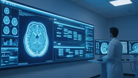

The global medical community continues to struggle with the staggering reality that brain tumors contribute to approximately a quarter of a million deaths annually, necessitating a radical shift in how clinicians approach magnetic resonance imaging analysis. Current diagnostic protocols often face bottlenecks due to the sheer volume of scans and the inherent complexity of differentiating between various pathological masses. Researchers at the University of Sharjah have recently introduced a sophisticated, multi-layered artificial intelligence framework designed to revolutionize neuro-oncological imaging by moving beyond standard deep learning models toward more complex, dual-stage architectures. This transition marks a significant milestone in medical technology, as it addresses the intricate nature of brain tumor diagnostics through a refined, binary-to-multiclass workflow. By focusing on this tiered approach, the team provides a more reliable tool for medical professionals who are increasingly burdened by high-volume screening demands and complex cases that require rapid and precise identification of specific tumor types.
The Mechanics of a Divided Diagnostic Pipeline
The proposed system functions by emulating the methodical logical workflow of a human radiologist, operating through two distinct phases to optimize both clarity and speed in a high-pressure clinical environment. Rather than attempting to solve every diagnostic variable in a single pass, the framework breaks the problem down into manageable components that ensure higher precision at each level of the pipeline. This modular approach allows for specific optimizations that a monolithic neural network would typically struggle to achieve, particularly when dealing with the subtle edge cases found in early-stage tumor growth. By creating a clear separation of concerns, the system maintains a high degree of reliability while remaining computationally efficient enough for integration into existing hospital infrastructures. This structural design offers a more intuitive model for clinicians to interact with, as the AI’s decision-making process mirrors the step-by-step diagnostic reasoning used today.
The First Stage: Binary Screening and Detection
The first stage serves as a critical binary gatekeeper, analyzing incoming MRI scans to determine whether a tumor is present or absent within the brain tissue. Through extensive comparative analysis, the research team identified the Vision Transformer as the most effective model for this initial screening task, allowing for high-accuracy identification of healthy versus pathological tissue in a high-traffic environment. Unlike traditional convolutional approaches that focus on local pixels, the Vision Transformer excels at capturing global dependencies, ensuring that even dispersed or unusually shaped anomalies are not overlooked. This preliminary layer acts as a high-speed filter, ensuring that medical resources are not wasted on negative scans while allowing radiologists to prioritize patients who require immediate surgical or therapeutic intervention. By maintaining a high sensitivity rate, the system provides a safety net that captures potential pathologies before they are passed to the classification stage.
The Second Stage: Multi-Class Categorization
Once the presence of a mass is confirmed, the system advances to a detailed multi-class classification stage to categorize the tumor into one of three primary types: glioma, meningioma, or pituitary tumors. To handle this more difficult task, the researchers deployed a hybrid model that merges Convolutional Neural Networks with Long Short-Term Memory networks. This combination creates a more robust framework for specific pathological identification than a single-model approach could provide, as it leverages the spatial feature extraction of CNNs alongside the sequential data processing of LSTMs. This dual-model synergy allows the system to recognize subtle variations in tumor borders and internal textures that indicate a specific malignancy. The result is a highly specialized diagnostic tool that provides clinicians with a precise starting point for treatment planning, significantly reducing the margin for error and improving long-term outcomes for patients.
Technological Synergies and Methodological Rigor
The success of this system highlights a broader trend toward attention-enhanced hybrid architectures in medical AI, where models are trained to prioritize specific regions of interest within an image. While standard networks excel at identifying local spatial features like textures and edges, the integration of memory networks allows the model to process information sequentially, capturing deeper relationships across the various slices of an MRI scan. This technological synergy is vital for understanding the full context of a tumor’s growth pattern and its interaction with surrounding brain structures. By combining different neural architectures, the system overcomes the limitations inherent in any single approach, providing a comprehensive analysis that is both spatially aware and contextually grounded. This holistic view is necessary for modern neuro-oncology, where the relationship between a mass and adjacent neurological pathways can dictate the feasibility of any surgical intervention or therapy.
Advanced Features: Attention Mechanisms and Noise Filtering
The most critical component of the feature awareness layer is the attention mechanism, which permits the model to focus on the most informative areas of the scan while filtering out irrelevant background noise or healthy anatomical structures. This selective focus is essential when dealing with tumors that may have poorly defined boundaries or those that closely resemble healthy tissue in certain lighting conditions. By mimicking the human ability to zoom in on anomalies, the AI achieves a level of precision that surpasses older, less sophisticated models that often struggled with low-contrast images. This approach not only improves diagnostic accuracy but also provides a more nuanced understanding of how different features contribute to the final result. Furthermore, the attention mechanism helps to reduce the likelihood of false positives by teaching the model to ignore common artifacts found in MRI scans, such as movement blur or magnetic field inhomogeneities.
Data Standardization: Preprocessing and Normalization Protocols
Beyond architectural innovation, the study emphasizes a standardized preprocessing pipeline to ensure objective and fair results across different tests and diverse patient populations. Using a dataset of over 7,000 MRI images, the team applied unified protocols for resizing and intensity normalization to minimize external variables that might confuse the neural network. This meticulous preparation ensures that the AI focuses solely on the actual pathology rather than variations in image quality or lighting conditions that often occur across different hospital imaging machines. Standardizing the input data is a crucial step in preventing the model from developing biases based on the specific hardware used to capture the scan. By creating a level playing field, the researchers have ensured the system remains reliable even when deployed in clinical settings with varying technical resources. This dedication to methodological rigor is what allows the framework to achieve high benchmarks in both sensitivity and specificity.
Computational Efficiency: High-Speed Inference for Clinical Use
For any AI tool to be viable in a fast-paced hospital environment, computational speed is just as important as diagnostic accuracy, as delays can lead to increased patient anxiety and postponed treatments. The University of Sharjah team reported inference times of less than 22 milliseconds per stage, suggesting the system is capable of near real-time feedback for clinicians during the diagnostic process. This efficiency could significantly alleviate the heavy workload currently experienced by radiology departments, acting as a secondary screening tool that accelerates the time between the initial scan and the start of life-saving treatment. In a modern healthcare setting, having a system that can process thousands of images in a matter of seconds allows human doctors to focus their expertise on the most complex cases. Furthermore, the low computational cost of these models means they can be deployed on standard hospital hardware without the need for expensive or specialized supercomputing clusters.
Future Horizons: Strategic Integration and Clinical Trust
The implementation of this dual-stage framework established a clear path forward for the integration of high-speed artificial intelligence in neuro-oncology. By successfully partitioning the detection and classification tasks, the research demonstrated that specialized hybrid models outperformed general-purpose architectures in both accuracy and efficiency. Moving forward, the focus shifted toward incorporating multi-modal data, such as genetic markers and patient history, to further refine the predictive capabilities of the system. Clinicians were encouraged to adopt these tools as collaborative assistants, using the AI to flag urgent cases while providing the human oversight necessary for patient care. The development of explainable AI modules became a priority to ensure that every diagnostic suggestion remained rooted in observable radiological features. Ultimately, the study proved that a structured, multi-layered approach to machine learning provided the necessary reliability to support critical medical decisions.