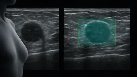

Advanced computational models are fundamentally reshaping oncological diagnostics by integrating sophisticated temporal-guided networks directly into clinical breast cancer workflows. This technological evolution marks a significant departure from traditional methods that rely heavily on the subjective interpretation of static, two-dimensional images. While ultrasound remains a cornerstone of medical imaging due to its cost-effectiveness and lack of ionizing radiation, it has long been plagued by high inter-observer variability, particularly when examining complex lesions. The recent introduction of a temporal-guided, multi-scale, multi-instance learning network, known as TGMIL, seeks to mitigate these challenges by utilizing the full depth of information contained within continuous ultrasound video clips. This paradigm shift moves away from looking at isolated snapshots of a potential malignancy and instead considers the entire dynamic narrative of the scan, providing a more holistic and reliable assessment of suspicious findings categorized under the Breast Imaging-Reporting and Data System (BI-RADS).
By focusing on the temporal continuity of a scan, the TGMIL system addresses the specific diagnostic hurdles encountered in regions with high breast tissue density, where standard mammography often fails to provide sufficient clarity. In these populations, ultrasound is the primary diagnostic tool, yet its success is intrinsically linked to the skill and experience of the sonographer performing the exam. The transition toward automated video analysis allows for a standardized evaluation process that identifies subtle morphological changes that might be missed during a live examination. This approach not only enhances the accuracy of initial screenings but also serves to reduce the number of false positives that lead to invasive and often unnecessary biopsies. The integration of such advanced systems into the daily routine of oncological departments represents a critical step in standardizing patient care and ensuring that diagnostic outcomes are no longer entirely dependent on the individual expertise of the practitioner.
Replicating Radiologist Expertise: The Power of Dynamic Analysis
Unlike many conventional artificial intelligence models that utilize “static learning” by analyzing a single, hand-picked representative frame from an ultrasound study, the TGMIL architecture was designed to mimic the actual cognitive process of a human expert. A radiologist does not form a final opinion based on a single image; rather, they move the probe across the skin, observing how the lesion’s internal echoes, margins, and relationship with surrounding tissues change in real-time. This dynamic observation allows them to distinguish between benign structures that might appear suspicious from one angle and true malignancies that maintain a consistent, irregular appearance throughout the scan. The TGMIL model replicates this by treating an entire ultrasound video as a single data unit, capturing the spatial-temporal evolution of the lesion as the imaging probe traverses the anatomy.
To achieve this level of sophistication, the network employs a Multi-Instance Learning (MIL) framework, which effectively treats a video as a “bag” containing multiple “instances” or frames. Each frame is processed through a convolutional backbone to extract visual features, while a temporal-guided component meticulously records the sequence and positional context of each image. This allows the system to understand the context of what it is seeing, identifying how a tumor’s appearance fluctuates throughout the examination. Furthermore, an integrated attention module acts as a sophisticated digital filter, automatically identifying which frames hold the most diagnostic weight. By prioritizing clear, representative segments of the video and suppressing noise or blurred frames caused by rapid probe movement, the model generates a comprehensive and highly accurate risk profile for the lesion in question.
Methodology and Comparative Performance: Validating the Model
The efficacy of the TGMIL system was established through a comprehensive multicenter study that analyzed 532 breast lesions across four distinct hospital settings, concluding its initial data collection phase in the current year. This diverse dataset allowed researchers to test the model’s robustness against varying equipment and sonography styles, ensuring that the results were not limited to a single institution. Each case included in the study was backed by definitive pathological findings obtained through biopsy or surgery, providing a solid “ground truth” for the AI to learn from. During the preprocessing stage, the researchers carefully removed all non-medical overlays, such as text and black borders, to prevent the algorithm from picking up irrelevant patterns. This rigorous approach ensured that the AI focused entirely on the physiological characteristics of the breast tissue.
In direct comparison with other leading diagnostic models, TGMIL showed a remarkable ability to balance sensitivity and precision, which is often the primary failure point for medical AI. In an independent test set of 198 lesions, the model achieved an Area Under the Receiver Operating Characteristic Curve (AUC) of 0.824, significantly outperforming traditional video classification architectures like 3D-CNN. While some newer static image models showed high sensitivity, they often suffered from low precision, frequently flagging benign cysts or fibroadenomas as malignant. TGMIL, however, maintained a sensitivity of 0.966 while keeping precision at 0.836, demonstrating that it could reliably identify cancer without causing a surge in “false alarms.” This balance is essential for clinical adoption, as it ensures that high-risk patients are correctly identified while minimizing the psychological and physical burden of unnecessary secondary testing on healthy individuals.
Impact on Sonographers: Bridging the Experience Gap
One of the most significant discoveries from this research involved a reader study designed to measure how the AI system influenced the decisions of clinicians with varying levels of experience. Sonographers were categorized into three groups: junior staff with under three years of experience, attending physicians with four to nine years, and senior experts with a decade or more in the field. For junior practitioners, the results were nothing short of transformative. Without the assistance of the AI, their diagnostic accuracy was recorded at approximately 66.7%; however, when they used the TGMIL system as a “second set of eyes,” their accuracy surged to 89.2%. This dramatic improvement suggests that the AI can effectively act as a tutor or safety net, providing less-experienced doctors with the specialized insight typically only acquired through years of high-volume clinical practice.
The interaction between the AI and senior experts, however, revealed a more complex relationship within the diagnostic workflow. These veteran radiologists already performed at an exceptionally high level, with an unaided accuracy that frequently matched or exceeded the standalone capabilities of the TGMIL model. Interestingly, when these experts were presented with AI suggestions that contradicted their initial findings, their performance occasionally dipped, a phenomenon known as automation bias or cognitive interference. This indicated that while the system was an invaluable asset for upskilling junior and mid-level staff, its utility for senior specialists was more limited. It highlighted the need for careful implementation strategies that allow experts to use the AI as a supplementary tool without overriding their seasoned clinical intuition, which remains a vital component of complex medical decision-making.
Implementation Strategies: Democratizing Specialized Oncological Care
A defining advantage of the TGMIL system is its remarkable computational efficiency and accessibility, as it was developed to function on consumer-grade hardware like the NVIDIA RTX 3090. This design choice was deliberate, ensuring that the technology could be integrated into existing hospital workstations without requiring massive investments in server infrastructure or specialized data centers. This portability is key to democratizing high-level care, particularly in rural or under-resourced medical facilities where access to senior breast imaging specialists is often restricted. By providing real-time feedback during a patient’s initial scan, the system enabled clinicians to make more informed decisions on the spot, potentially reducing the time between the initial discovery of a lesion and the commencement of a treatment plan.
In the final assessment of the project’s impact, stakeholders emphasized that the successful deployment of video-based AI required a shift in how medical data was standardized across different healthcare networks. The research team identified that while TGMIL performed admirably in a controlled multicenter study, real-world application demanded continuous updates to account for the evolving nature of ultrasound technology and a wider variety of rare lesion types. Clinicians who participated in the study suggested that future iterations should focus on integrating multi-modal data, such as patient history and elastography, to further refine the model’s predictive power. By establishing these actionable steps, the medical community moved closer to a collaborative diagnostic environment where machine precision and human expertise combined to ensure that no malignancy was overlooked, while simultaneously protecting patients from the strain of over-diagnosis.