
The insidious nature of heart valve disease often allows it to progress undetected until the cardiovascular system reaches a critical breaking point, making early and precise quantification of mineral deposits a cornerstone of modern cardiology. The innovative computational architecture for valve analysis involves slicing volumetric data into eleven contiguous short-axis frames for comprehensive spatial evaluation. This methodology addresses a long-standing gap in clinical diagnostics, where the gold standard for assessing aortic valve calcification has traditionally relied on ionizing radiation and expensive computed tomography scans. By pivoting toward an artificial intelligence-driven pipeline that utilizes three-dimensional transesophageal echocardiography, medical professionals are now exploring a safer, more accessible alternative. This pilot study, focusing on the feasibility and technical accuracy of ultrasound-based calcium scoring, signals a significant departure from the reliance on Agatston scores derived from CT imaging, offering a radiation-free pathway for patients with moderate to severe aortic stenosis who require frequent monitoring.
Technological Evolution in Imaging
Enhancing Ultrasound Precision: From Visuals to Data
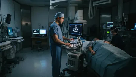
The shift toward three-dimensional transesophageal echocardiography, or 3D TEE, represents a major physical and technical upgrade over traditional transthoracic methods. By positioning the ultrasound probe within the esophagus, clinicians can bypass the physical barriers of the rib cage and lung tissue, which often create significant acoustic interference. This anatomical proximity allows for the use of much higher acoustic frequencies, resulting in a dramatic improvement in spatial resolution. Historically, cardiologists had to rely on a qualitative assessment of valve health, often grading calcium burden as mild, moderate, or severe based on the subjective brightness of the leaflets on a screen. This new computational approach removes that human variability by applying an automated heuristic that converts visual echoes into a reproducible numerical dataset, effectively standardizing how valve health is recorded across different clinical settings and hardware manufacturers.
Beyond the immediate physical advantages of probe placement, the integration of 3D volumetric analysis allows for a more holistic view of the cardiac architecture. Standard ultrasound often captures only a two-dimensional “slice,” which can easily miss localized clusters of calcification that might significantly impact the valve’s mechanical function. By utilizing 3D datasets, the AI can analyze the entire volume of the aortic valve apparatus, ensuring that no mineralized deposits are overlooked. This transition from a “visual impression” to a data-heavy quantification process is essential for precision medicine in 2026. As heart disease management becomes more personalized, having a granular, pixel-based metric of disease progression allows for better-timed surgical interventions, ultimately reducing the risk of heart failure in patients who previously sat in a diagnostic gray area between different imaging modalities.
Study Demographics: Validating the Diagnostic Baseline
The validation of this AI-driven approach was conducted through a rigorous prospective pilot study involving twenty-three patients currently diagnosed with varying degrees of aortic stenosis. With a median age of seventy-six and a slight male predominance, the cohort represented the high-risk demographic most commonly affected by degenerative valve disease. Most participants suffered from severe stenosis, characterized by significant narrowing of the valve orifice and elevated pressure gradients. To provide a scientific anchor for the algorithm’s performance, researchers included healthy control subjects with non-calcified valves. This inclusion was critical for establishing a “zero-baseline,” training the software to recognize the supple, thin nature of healthy tissue and preventing it from generating false-positive signals that could lead to unnecessary clinical concern or misdiagnosis.
Consistency in the timing of comparative diagnostics was a central pillar of the study’s design to ensure that any findings were a true reflection of the technology’s capabilities rather than a result of disease progression. Each patient underwent both the experimental 3D TEE scan and a traditional cardiac CT scan within a tight sixty-five-day window. In the context of aortic stenosis, this timeframe is considered stable enough to assume that the level of calcification would not have changed significantly between the two procedures. By maintaining this strict protocol, the research team could directly correlate the AI’s ultrasound-derived pixel counts with the established Agatston scores provided by CT imaging. This comparison serves as the ultimate benchmark for the software, proving that ultrasound can provide the same level of diagnostic insight without the associated risks of radiation or contrast-induced kidney stress.
The AI Computational Pipeline
Multi-Step Data Processing: Reconstructing the Volumetric View
The computational heavy lifting begins during the diastolic phase of the cardiac cycle, a moment when the aortic valve is closed and at its most physically stable point for imaging. The AI pipeline utilizes a multiplanar reconstruction technique that takes the raw 3D TEE volumetric data and slices it into eleven contiguous short-axis frames. Each of these slices is precisely 1.5 millimeters thick, a depth chosen to ensure that the entire vertical span of the valve and its surrounding annulus is captured in high detail. This systematic slicing prevents the “skipping” of data that often occurs in traditional 2D imaging, providing a comprehensive map of where mineralized deposits are located. This phase is entirely digital, transforming a single complex volume into a stack of high-resolution images ready for deep-level pixel analysis.
A critical human-in-the-loop component remains essential during this stage of the process to ensure anatomical accuracy. Clinicians manually define the region of interest, a step that guides the AI to focus exclusively on the aortic valve and its supporting structures. This is a vital safeguard, as the heart is a dense environment where calcium can also accumulate in the nearby coronary arteries or the mitral valve. By explicitly delineating the boundaries of the aortic apparatus, the software can ignore these adjacent calcifications, ensuring that the final score is a pure reflection of the aortic valve’s health. This focused approach reduces the “noise” in the data, allowing the algorithm to operate with a high degree of specificity that is required for making critical decisions about valve replacement or alternative transcatheter therapies.
From Pixels to Scores: Quantification Through Binarization
Once the images are reconstructed and the region of interest is set, the software performs a complex conversion of the standard grayscale ultrasound signal into a Hue, Saturation, and Value (HSV) color space. This conversion is the first step in a sophisticated computer vision routine that uses edge detection and contouring to isolate the valve leaflets from the surrounding blood pool and soft tissue. By analyzing the unique signal characteristics of calcified deposits—which reflect ultrasound waves much more intensely than flexible tissue—the algorithm can pinpoint exactly where mineralization has occurred. This transition from a simple grayscale image to a multi-dimensional color space allows the software to differentiate between various tissue densities with a level of precision that far exceeds the capabilities of the human eye.
The final stage of the quantification process involves a digital binarization technique that converts the image into a map of absolute values. The AI applies a specific signal intensity threshold to every individual pixel within the slices; those that exceed the threshold are classified as “calcium-positive” and represented as white pixels, while everything below that threshold is discarded as black space. The total “AI-derived calcium score” is then calculated by summing every white pixel across all eleven frames. This provides a direct, objective numerical value that represents the total volume of calcification. This pixel-based scoring system effectively creates a new language for ultrasound diagnostics, allowing clinicians to track the growth of mineral deposits over time with a degree of mathematical certainty that was previously the exclusive domain of radiographic imaging.
Analyzing Clinical Correlations
Statistical Alignment: Evaluating Ultrasound against Computed Tomography
The success of the AI-driven ultrasound score hinges on its ability to mirror the clinical insights provided by the traditional CT Agatston score, and the study’s results demonstrated a strong statistical alignment between the two. Initial analysis showed a significant positive relationship with a Spearman rank correlation coefficient of 0.55, indicating that as the CT-detected calcium increased, the ultrasound-derived pixel count followed a similar trajectory. This correlation became even more pronounced during a sensitivity analysis that accounted for extreme outliers. In one specific case, a patient exhibited an exceptionally high Agatston score exceeding 10,000, which created significant acoustic shadowing that hindered the ultrasound’s performance. When this outlier was removed, the correlation coefficient rose to 0.65, proving that for the vast majority of patients in a standard clinical setting, the ultrasound metric is a highly reliable surrogate for CT data.
This statistical consensus suggests that 3D TEE, when enhanced by AI, can effectively replicate the diagnostic utility of radiation-based imaging for risk stratification. The ability to achieve these results is a testament to the robustness of the signal processing algorithms used to filter out noise while preserving the integrity of the calcium signal. For clinicians, this means that the ultrasound data they already collect for surgical planning can now provide a secondary layer of objective risk assessment. Instead of relying on a separate, potentially hazardous CT scan to confirm the severity of a patient’s condition, the AI provides a comprehensive picture of the mineral burden directly from the echocardiogram. This consolidation of diagnostic data not only streamlines the clinical workflow but also ensures that patients receive a more holistic evaluation during a single, safer imaging session.
Performance Metrics: Benchmarking Accuracy for High-Risk Care
The diagnostic power of the AI system was further validated through Receiver Operating Characteristic analysis, which measures how effectively a test can distinguish between different levels of disease severity. The AI metric achieved an Area Under the Curve (AUC) of 0.87 for detecting severe calcification, a score that indicates high diagnostic accuracy and reliability. By establishing a specific cutoff value of approximately 68,813 pixels, the system demonstrated nearly 90% sensitivity, meaning it was highly effective at identifying patients who truly suffered from severe mineral deposits. This level of precision is critical in 2026, where the timing of valve replacement is increasingly dictated by the objective burden of calcium rather than just the patient’s symptoms, which can often be vague or masked by other comorbidities.
Beyond simply identifying severe cases, the AI model maintained a strong AUC of 0.82 when differentiating the most extreme, high-risk tiers of the disease. This ability to categorize patients into different risk brackets is essential for long-term management, as those with rapidly increasing pixel scores may require more frequent follow-ups or a move toward earlier intervention. The results confirmed that the software does not just provide a binary “yes or no” regarding the presence of calcium but offers a nuanced spectrum of data that aligns closely with established clinical benchmarks. By providing a radiation-free method to achieve this level of detail, the technology addresses a critical need in cardiovascular medicine, offering a way to monitor the most vulnerable patients without the cumulative risks associated with repeated CT scans.
Implementation and Future Care: Expanding the Diagnostic Horizon
The pilot study concluded that the integration of AI with 3D transesophageal echocardiography significantly improved the objective quantification of aortic valve calcification, providing a reliable alternative to radiation-based imaging. Researchers successfully demonstrated that a pixel-based metric could serve as a surrogate for the Agatston score, particularly in patients with moderate to severe stenosis. This finding is expected to catalyze a shift toward “opportunistic” diagnostics, where high-value data is extracted from standard clinical procedures. By leveraging software to gain CT-level insights from ultrasound, healthcare systems can reduce the financial and logistical burdens associated with multi-modality imaging. The study established a clear proof of concept, showing that computational analysis can transform subjective visual interpretations into actionable, reproducible medical data.
Moving forward, the primary focus will be on the transition from semi-automated workflows to fully autonomous diagnostic systems. Actionable next steps include the development and integration of deep learning models designed for autonomous valve segmentation, which will remove the need for manual region-of-interest definition by clinicians. This advancement will likely increase the speed of analysis and further reduce the potential for human error, making the technology more suitable for high-volume clinical environments. Additionally, the medical community should prioritize multicenter trials with larger, more diverse patient populations to refine the algorithm’s performance across different hardware platforms. As these AI pipelines become more standardized, they will offer a safer, more sustainable pathway for the serial monitoring of valvular disease, ensuring that interventions are timed with the highest possible precision.