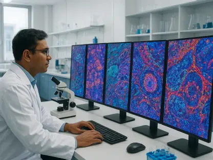

Automating the volumetric measurement of meningiomas requires navigating the intricate boundaries of tumors that grow along brain membranes, making standard geometric analysis insufficient for accurate tracking. For years, clinicians have struggled with the limitations of two-dimensional imaging, which often fails to capture the asymmetrical growth patterns of these common primary brain tumors. Because meningiomas expand along the curved surfaces of the dura mater, their true burden is frequently underestimated by conventional diameter-based assessments. The current medical landscape demands a transition toward three-dimensional volumetric analysis, yet the manual labor required for such precision is unsustainable in high-volume hospital settings. This necessity has driven the development of automated solutions that can assist neuroradiologists without sacrificing accuracy. However, the adoption of artificial intelligence has been hampered by a lack of transparency, leading to a critical need for systems that not only provide data but also communicate their level of confidence in every measurement.
Bridging the Gap With Evidential Deep Learning
Technical Innovations: Uncertainty Mapping in Neuro-Oncology
At the heart of this technological advancement is a sophisticated framework known as Evidential Deep Learning, which distinguishes itself from conventional neural networks by acknowledging its own limitations. Standard artificial intelligence models typically offer a definitive output, which can be dangerously overconfident when presented with ambiguous imaging data. This “black box” nature has long been a barrier to clinical adoption, as doctors are understandably hesitant to rely on a system that cannot explain its reasoning or admit when a case is difficult. By contrast, the University of California, San Francisco researchers implemented an architecture that quantifies uncertainty directly within the learning process. This allows the model to categorize information not just by what it sees, but by how reliable those observations are based on the training it has received. This move toward self-aware algorithms is a fundamental departure from the rigid models of the early 2020s.
The most visible manifestation of this approach is the creation of uncertainty maps, which act as a secondary layer of information for the interpreting radiologist. These digital overlays highlight specific regions where the algorithm is less confident about the boundary between a tumor and the surrounding brain tissue. Instead of forcing the physician to hunt for potential errors, the system proactively flags these zones, effectively guiding the human expert to the most complex areas of the scan. This synergy between machine and human ensures that the radiologist’s time is spent where it is most needed—resolving ambiguities rather than performing repetitive manual segmentation. By surfacing these doubts, the tool transforms from a mysterious automated process into a transparent assistant that actively communicates its internal logic. This transparency is crucial for building the necessary trust to integrate such advanced diagnostic tools into the fast-paced environment of a modern hospital.
Model Validation: Performance Across Diverse Medical Datasets
To ensure the model could survive the rigors of a real-world clinical setting, the UCSF team utilized an extensive dataset comprising over 1,600 MRIs, far exceeding the scale of typical pilot studies. A central challenge in this process was the inclusion of post-operative scans, which are notoriously difficult for automated systems to navigate. After a patient undergoes surgery, the resulting inflammation, scar tissue, and surgical debris can create visual signatures that closely mimic the appearance of a recurring tumor. Most existing algorithms struggle with these artifacts, leading to high rates of false positives that can cause unnecessary alarm for both patients and clinicians. By training the model on these complicated cases, the researchers ensured that the technology remained robust even when the anatomy was distorted by previous interventions. This rigorous training protocol established a baseline for accuracy that few other tools have achieved in such a diverse patient population.
In addition to the sheer volume of data, the researchers employed ensemble architectures to further bolster the reliability of the system’s outputs. This method involves running multiple independent models simultaneously and requiring them to reach a consensus, much like a panel of doctors discussing a complex case. If the different models disagree on a specific section of the scan, that area is marked with high uncertainty, alerting the user to a potential discrepancy. This “wisdom of the crowd” approach significantly reduces the likelihood of systemic bias or fluke errors that might plague a single neural network. Furthermore, the framework was validated against external datasets from various hospitals to ensure it was not overfitted to specific imaging hardware. This cross-institutional testing proved that the model maintains its predictive power across different MRI scanner brands and magnetic field strengths, making it a versatile solution for global medical facilities.
Establishing Accuracy and Clinical Alignment
Enhanced Precision: Improving Interpretability in Radiologic Reviews
The clinical validation phase of the study demonstrated that the AI achieved an impressive level of spatial agreement with the assessments of senior neuroradiologists. This is particularly significant because 3D volumetric analysis has consistently proven more sensitive than traditional 2D metrics for detecting subtle changes in tumor burden. While 2D measurements only look at the widest points of a growth, the 3D model evaluates the entire mass, providing a far more accurate representation of the disease’s trajectory. This level of granularity allows for the detection of growth that might otherwise be dismissed as stable under older imaging standards. By providing a precise numerical value for the tumor volume, the tool removes much of the variability that occurs when different doctors interpret the same set of images. This standardization is a massive step forward in ensuring that every patient receives a consistent level of care, regardless of which radiologist happens to be on duty.
Another vital finding was the high degree of calibration between the model’s reported uncertainty and its actual error rate. In clinical terms, this means that when the system expressed low confidence in a particular area, there was a statistically higher probability that its initial assessment was incorrect. This alignment mirrors the way human experts process information; a radiologist knows when they are looking at a particularly difficult case and proceeds with extra caution. By mimicking this human trait, the AI provides a reliable metric for its own fallibility, which is perhaps more important than the raw accuracy score itself. When the uncertainty maps indicate a high degree of doubt, the system essentially “asks” for human intervention, ensuring that the final diagnosis is a product of both machine speed and human experience. This calibration prevents the dangerous overreliance on automation that has characterized earlier iterations of artificial intelligence in the medical field.
Future Perspectives: The Evolution of AI-Driven Oncology care
Looking ahead, the successful deployment of this framework has paved the way for larger, multi-center trials that will further refine the algorithm’s performance. Researchers have planned to explore multi-rater annotations, which involves comparing the AI’s uncertainty scores with the natural variability found among a diverse group of human experts. This will help determine if the AI’s “doubt” aligns with the areas where even the most experienced doctors tend to disagree. Additionally, there is significant interest in expanding this uncertainty-aware architecture to other types of brain tumors and even different organ systems. By broadening the scope of the training data, the technology could eventually become a universal standard for volumetric monitoring across the entire field of radiology. These future developments are expected to focus on real-time integration into hospital systems, making the tool a seamless part of the daily routine for radiologists around the world.
The development of this uncertainty-aware model represented a pivotal moment in the evolution of neuro-oncology, as it successfully addressed the critical issue of trust in medical automation. By prioritizing transparency and safety, the UCSF team provided a blueprint for how artificial intelligence could be safely integrated into the most sensitive areas of patient care. Healthcare organizations were encouraged to adopt these standards of explainability to ensure that machine efficiency never came at the cost of clinical accuracy. Moving forward, the focus shifted toward establishing clear regulatory guidelines for the use of uncertainty metrics in diagnostic software. This research effectively proved that a machine’s ability to admit what it did not know was just as valuable as its ability to identify what it did. As these systems became more common, they fundamentally altered the landscape of cancer monitoring, ensuring that the synergy between human intuition and algorithmic precision remained the gold standard.