
Standard ADC measurements frequently result in unnecessary biopsies for benign findings due to their inability to resolve the internal heterogeneity of complex breast lesions. Modern oncology is currently undergoing a significant shift as clinical researchers move beyond the standard limitations of conventional imaging to unlock deep, hidden data sets contained within breast tumors. A breakthrough study recently conducted by a collaboration between Tsinghua University and Fudan University Shanghai Cancer Center, and subsequently published in the journal BMC Medical Imaging, has highlighted a new frontier for diagnostic accuracy in the medical field. By merging high-dimensional data extraction with advanced microstructural mapping, this recent research suggests that the medical community can finally move past the era of subjective interpretation toward a more precise, data-driven approach for identifying malignancies. The primary challenge in current breast cancer screening protocols stems from a heavy reliance on the average values of the Apparent Diffusion Coefficient.
Bridging the Gap: Advanced Imaging Techniques
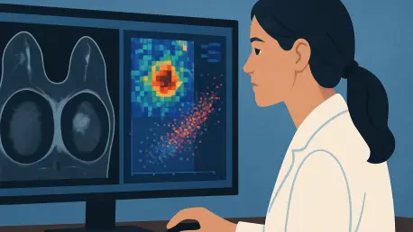
To address these systemic shortcomings, the study introduced MR cytometry, a sophisticated technique that utilizes multi-diffusion-time acquisitions through specialized MRI sequences. Unlike traditional scans that typically provide a static snapshot of tissue, MR cytometry probes water diffusion across various scales, creating a robust spatial visualization of tissue architecture. This specific methodology allows for a detailed mapping of cellular density and organization, providing a much richer foundation for clinical analysis than the simplified numerical averages used in conventional hospital settings. By utilizing both pulsed gradient spin-echo and oscillating gradient spin-echo sequences, the researchers successfully captured the complex nuances of the tumor environment. This technological leap allows clinicians to visualize how water molecules interact with cellular membranes at a microscopic level, offering a level of detail that was previously inaccessible through standard imaging protocols, thereby laying the groundwork for a more nuanced diagnosis.
Complementing these detailed mappings is the field of radiomics, which employs machine learning algorithms to mine medical images for hundreds of unique pixel-level features. These features, which encompass shape, first-order intensity statistics, and highly complex texture patterns, are often entirely invisible to the naked human eye during a routine radiological review. By applying radiomic analysis specifically to MR cytometry mappings, the research team was able to identify subtle signatures of malignancy that would otherwise remain hidden within the noise of standard scans. This process essentially transforms standard medical images into large, searchable datasets filled with biological information. Rather than looking for a single visual cue, the AI scans for mathematical relationships between pixels that correlate with specific pathological states. This multi-layered approach ensures that the diagnostic process is not just about identifying a mass, but about understanding the very fabric of the tissue being examined in a quantitative manner.
Validating Accuracy: Rigorous Methodological Standards
The strength and validity of this study lie in its prospective, two-center design, which involved 221 patients with pathologically confirmed breast lesions to ensure that the findings were not limited to a specific facility or demographic. The researchers employed a sophisticated and highly controlled machine-learning pipeline, utilizing dimensionality reduction techniques like Principal Component Analysis and the Least Absolute Shrinkage and Selection Operator to prevent the models from overfitting. This rigorous approach ensured that the artificial intelligence was learning genuine diagnostic patterns rather than merely memorizing coincidental data artifacts or background noise from the imaging equipment. By validating the results against an external test cohort, the team demonstrated that their methodology was reproducible and robust enough for potential real-world clinical applications. This level of methodological rigor is essential for building trust among medical professionals who are often skeptical of black-box AI solutions that lack transparency in their training.
The numerical results of this methodology were quite striking, demonstrating that the combined radiomics and MR cytometry approach significantly outperformed traditional diagnostic methods across every tested metric. While standard Apparent Diffusion Coefficient models achieved a respectable level of accuracy, the integration of radiomic features from MR cytometry maps pushed the Area Under the Curve to an impressive 0.914. When both traditional ADC and advanced cytometry features were combined into a single diagnostic model, the predictive power reached a peak AUC of 0.935 in the training phase. This finding is particularly significant because it suggests that the richness and quality of the underlying data, rather than the raw complexity of the machine learning architecture, were the primary drivers of diagnostic success. Even relatively simple logistic regression classifiers performed exceptionally well when fed the high-quality mappings produced by the cytometry process, proving that better data leads to better clinical outcomes without needing overly convoluted AI structures.
Clinical Transformation: Future Insights and Outcomes
This research signals a broader movement within the medical community toward the concept of non-invasive virtual biopsies, where the entire volume of a tumor is analyzed without the need for needles or surgical intervention. Traditional biopsies only sample a very small fraction of a lesion, which often fails to reflect its overall character due to the inherent internal diversity of cancer cells. In contrast, the quantitative mining of MRI data provides a comprehensive, three-dimensional view of the entire tumor, offering a safer and more holistic alternative for evaluating suspicious findings during screening. This shift toward whole-tumor analysis is critical because it captures the most aggressive portions of a malignancy that might be missed by a blind needle stick. By providing a global map of the tumor’s microstructural environment, clinicians can make more informed decisions about treatment pathways and surgical planning, ensuring that the chosen intervention is tailored to the specific biological profile of the patient’s individual disease state.
The integration of these advanced imaging tools successfully redefined the potential of breast cancer diagnostics by providing a clear roadmap for more efficient and accurate patient triaging. Hospital administrators and radiology departments sought to implement these methods through seamless software updates, as the technology utilized existing MRI hardware without requiring additional contrast agents. This approach offered a practical solution for reducing the number of unnecessary biopsies, thereby alleviating patient anxiety and lowering overall healthcare expenditures. While future investigations focused on expanding the dataset to include more diverse populations and a higher ratio of benign cases, the initial breakthroughs established a new standard for non-invasive care. Clinical teams began to rely on these high-dimensional mappings to distinguish between stable lesions and those requiring immediate intervention. Ultimately, the transition to radiomics-based cytometry proved that deep data mining was the key to unlocking the full potential of medical imaging, ensuring that every pixel contributed to a more certain and life-saving diagnosis for patients.