
Clinical validation of the COMBINE-CT platform marks a major milestone in the effort to create a seamless, automated workflow for heart disease. This transition represents a fundamental shift in how medical professionals approach the diagnosis and management of the world’s leading cause of mortality. In the current landscape of 2026, the traditional reliance on manual interpretation is giving way to sophisticated computational models that process vast datasets in seconds. For decades, the bottleneck in cardiac care was the time-consuming process of human review, which often led to delays in treatment or the over-utilization of invasive procedures. By integrating advanced machine learning directly into imaging hardware, clinicians are now seeing a level of detail in coronary artery assessments that was previously unattainable. This evolution is about providing a more nuanced understanding of patient-specific risk factors through high-fidelity digital twins of the human heart.
Evolution of Cardiac Precision: Advancing Diagnostic Imaging
The integration of automated fractional flow reserve (FFR-CT) and plaque characterization software has fundamentally changed the criteria for surgical intervention. Rather than relying solely on the narrowing of an artery, physicians now utilize AI to simulate blood flow dynamics under various stress conditions. This physiological assessment, performed entirely non-invasively, allows for a more accurate determination of which lesions are truly causing ischemia and which can be managed safely with medication. Furthermore, AI-driven tools are now capable of distinguishing between stable calcified plaques and high-risk vulnerable plaques that are prone to rupture. By identifying these lipid-rich features early, the medical community has successfully pivoted toward a preventive model of care. This capability ensures that high-risk patients receive aggressive intervention before a cardiac event occurs, while low-risk individuals are spared the physical and financial burdens of any unnecessary procedures.
Moving beyond simple detection, the current generation of AI-enabled scanners operates as a definitive diagnostic tool rather than a mere screening mechanism. This shift has significantly reduced the diagnostic odyssey for patients presenting with chest pain, as a single CT scan now provides the comprehensive data previously requiring multiple stress tests. By centralizing data processing through unified platforms, hospitals are seeing a drastic reduction in inter-observer variability, which has historically been a challenge in cardiac imaging. When every scan is analyzed through the same rigorous algorithmic lens, the consistency of care improves across diverse demographics and regions. This standardization is critical for large-scale healthcare networks seeking to optimize patient outcomes while managing operational costs. The ability to generate a high-confidence diagnosis within minutes has redefined the standard of care, making rapid-response cardiology a reality in both urban and rural environments.
Strategic Implementation: Integrating Automation into Clinical Practice
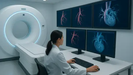
The practical implementation of these technologies involves a shift toward cloud-based infrastructure that supports real-time data sharing and longitudinal tracking of heart health. Hospitals that have adopted these systems from 2026 to the planned expansions in 2028 are finding that the biggest benefit is the reduction in cognitive load on specialized staff. Instead of spending hours mapping out 3D models of the coronary tree, radiologists now review pre-rendered, AI-annotated images that highlight areas of concern for human verification. This collaborative model, often referred to as human-in-the-loop AI, leverages the pattern recognition of software alongside the clinical judgment of experienced doctors. The result is a more resilient healthcare system where practitioners focus on patient communication and treatment planning rather than rote data entry. This streamlined approach also facilitates the integration of CT data with electronic health records, ensuring every member of a team has access to the insights.
Looking at the trajectory of cardiac medicine, health systems that embraced these automated platforms achieved a notable reduction in readmission rates and improved long-term survival statistics. To maintain this momentum, stakeholders focused on establishing robust data governance frameworks and investing in continuous training for the radiological workforce. The successful transition required a departure from siloed imaging departments toward an integrated multidisciplinary approach where AI served as the connective tissue between diagnostics and therapeutics. For those looking forward, the primary objective remained the optimization of these algorithms for diverse patient populations to eliminate diagnostic biases. By prioritizing the procurement of hardware with native AI integration and ensuring seamless interoperability, medical institutions secured their place at the forefront of modern cardiology. These strategic investments proved that technology can bridge the gap between complex digital data and life-saving clinical action.