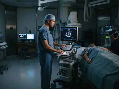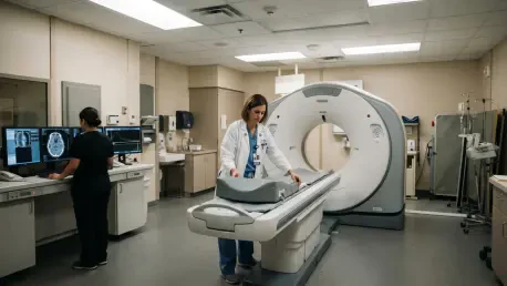

Modern radiology departments are exploring adaptive dosing strategies where real-time imaging feedback is used to adjust cancer treatments for individual patient responses. This shift marks the beginning of a new era where diagnostic imaging is no longer a passive observation but a dynamic driver of therapeutic intervention. The convergence of molecular biology, high-speed computing, and legislative mandates is dismantling the traditional silos that once separated radiologists from the direct care of their patients. As we navigate the landscape of 2026, the medical community is witnessing a fundamental redefinition of the specialist’s role, moving away from isolated image interpretation toward holistic, longitudinal involvement in the patient journey. This evolution is driven by the necessity to provide more personalized care in an increasingly complex oncological environment. By integrating advanced data analytics with highly targeted treatment modalities, practitioners are now equipped to offer insights that were previously obscured by the limitations of conventional technology. The result is a more resilient healthcare system where the imaging specialist serves as a central architect of the clinical strategy, ensuring that every diagnostic finding translates directly into an actionable and effective therapeutic step for the individual.
Impact of the 21st Century Cures Act
The 21st Century Cures Act has fundamentally altered the relationship between imaging specialists and those they serve by mandating immediate digital access to medical records. Historically, radiologists functioned as “doctors’ doctors,” with their reports intended primarily for referring physicians who would then filter and interpret the findings for the patient. However, the requirement for instantaneous transparency has shifted the dynamic, placing diagnostic reports directly into the hands of patients, often at the same moment the ordering physician receives them. This change has removed the traditional buffer and created a new urgency for clarity and accessibility in medical documentation. Patients are now more empowered than ever, frequently arriving at their clinical appointments having already read their own scans and formulated specific questions. Consequently, the radiology report is no longer a purely technical memorandum; it has become a primary communication tool that must balance clinical precision with a level of readability that respects the patient’s right to understand their own health status without unnecessary anxiety or confusion.
This shift toward real-time report access necessitates an evolution in communication models across all imaging subspecialties. Even when a physical face-to-face meeting does not occur, the radiology report now serves as a direct point of interaction between the specialist and the patient, acting as a narrative bridge in the care continuum. Imaging professionals are increasingly encouraged to adopt a mindset where they practice as if they are in the room with the patient, prioritizing holistic communication that anticipates and addresses potential patient concerns within the text of the report itself. This proactive approach is preparing the field for a future where patients may regularly seek direct consultations with radiologists to interpret complex findings or discuss the implications of advanced molecular imaging. By embracing this transparency, radiologists are strengthening their clinical relevance and demonstrating that their expertise extends far beyond the reading room. This transition from a back-end service to a front-facing clinical partner ensures that the patient remains at the center of the diagnostic process, fostered by a culture of openness and mutual understanding.
Advancements in Artificial Intelligence Adoption
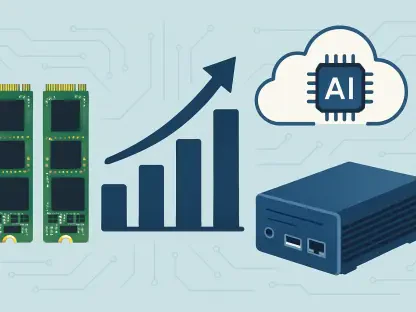
Artificial Intelligence has transitioned from a source of professional apprehension to a vital tool for clinical efficiency, particularly as radiology is fundamentally a digital field operating within sophisticated Picture Archiving and Communication Systems. Because the work of the radiologist is already embedded in a digital workflow, the integration of algorithmic tools for abnormality detection and triage has been a natural progression rather than a disruptive shock. Today, AI is being leveraged to standardize reporting across diverse healthcare systems, ensuring a consistent level of quality and accuracy regardless of the facility’s location or size. These tools act as a second set of eyes, reducing fatigue-related errors that can occur during high-volume shifts and allowing physicians to focus their cognitive energy on the most complex and nuanced cases. Furthermore, the automation of routine measurements and administrative tasks has streamlined the diagnostic process, enabling faster turnaround times for critical results and improving the overall throughput of busy imaging centers without sacrificing the depth of analysis required for precision medicine.
A particularly promising application of AI in the current clinical environment is the generation of plain language summaries within complex medical reports. By translating dense technical terminology and anatomical jargon into accessible information, AI helps bridge the gap between specialist knowledge and patient understanding, facilitating a more productive dialogue between patients and their providers. Looking forward, the next frontier in this technological evolution is the closure of the “outcomes gap” through predictive modeling. Rather than merely identifying the presence of a lesion, advanced algorithms are now being trained to integrate imaging data with electronic medical records and genomic profiles to forecast how a specific individual will respond to a particular treatment regimen. This shift from simple detection to sophisticated prediction allows for the optimization of therapy before it even begins, identifying patients who are likely to benefit from specific interventions while sparing others from ineffective treatments and their associated side effects. By moving toward this predictive paradigm, radiology is solidifying its role as a cornerstone of data-driven oncology.
The Revolutionary Potential of Theranostics
Theranostics represents the ultimate expression of precision medicine by combining “therapy” and “diagnostics” into a single, highly targeted approach that treats what it sees and sees what it treats. This process involves identifying specific proteins or receptors on the surface of cancer cells, such as the Prostate-Specific Membrane Antigen in prostate cancer, and using a diagnostic isotope to visualize the extent of the disease with high-resolution PET imaging. Once the presence and location of the target are confirmed, the diagnostic isotope is substituted for a therapeutic one, which delivers a localized dose of radiation directly to the malignancy. This “lock and key” mechanism allows for the eradication of microscopic disease that might be invisible to conventional scans, all while sparing the surrounding healthy tissue from the collateral damage typically associated with external beam radiation or systemic chemotherapy. The ability to verify the target prior to treatment ensures a high degree of confidence in the therapeutic strategy, minimizing the trial-and-error approach that has historically characterized much of oncology.
While theranostics has found significant initial success in treating prostate and neuroendocrine tumors, the potential for this platform extends to a wide array of other malignancies, including breast, lung, and pancreatic cancers. The future of this field lies in the implementation of adaptive dosing strategies, where the treatment is adjusted in real-time based on the patient’s unique physiological response as observed through interim imaging. This level of personalization allows for the optimization of the radioactive payload, ensuring that the dose is high enough to be effective but low enough to maintain the patient’s quality of life. Furthermore, the ability to combine these radioactive isotopes with other modern treatments, such as immunotherapy or specialized chemotherapy, positions theranostics as a versatile and indispensable tool in the multimodal fight against cancer. As clinical trials continue to expand the list of viable targets, the theranostic approach is becoming a standard pillar of care, offering hope to patients with advanced disease who previously had few options for effective management.
New Milestones in Targeted Isotopes
Innovation in the management of systemic disease is currently focused on the transition from traditional beta-emitting isotopes to more potent and localized alpha particles. These particles, which include isotopes such as Actinium-225, Terbium-161, and Lead-212, possess significantly greater mass and energy than their predecessors but travel a much shorter distance through human tissue. This physical characteristic is crucial because it allows for the delivery of an incredibly high-energy payload to a very localized area, effectively destroying the DNA of cancer cells within a range of only a few cell diameters. By limiting the radiation’s reach, alpha particles minimize the impact on adjacent healthy organs, which is particularly beneficial when treating tumors located near sensitive structures like the salivary glands or the bone marrow. This precision allows for a more aggressive treatment of the cancer while simultaneously improving the overall safety profile, leading to fewer side effects and a better quality of life for patients undergoing intensive therapy.
The significant influx of capital into alpha-particle research and development signals a high level of market and clinical confidence in the future of the theranostics platform. Pharmaceutical companies and research institutions are racing to develop new ligands that can carry these potent isotopes to a variety of different cancer markers, expanding the reach of targeted radiotherapy beyond its current boundaries. These technological milestones suggest that the field is moving toward a more nuanced and effective method of disease eradication that prioritizes biological specificity over broad-spectrum intervention. By focusing on these high-energy, short-range isotopes, researchers are achieving more durable responses in patients with metastatic disease who may have exhausted other conventional treatment options. The ongoing refinement of isotope production and delivery systems ensures that these life-saving therapies are becoming more widely available, transforming what was once a niche research interest into a mainstream clinical reality that is redefining the expectations for long-term cancer survival.
Embracing Technological Optimism over Fear
Despite recurring anxieties that the rapid advancement of Artificial Intelligence might eventually replace human physicians, historical parallels suggest that technology more often acts as a catalyst for growth and specialization rather than a replacement for professional expertise. The introduction of digital imaging and teleradiology in previous decades sparked similar fears of job loss and the commoditization of the field, yet these tools ultimately increased clinical efficiency and led to an even greater demand for highly trained radiologists. In the current environment, the focus has shifted toward using AI to fill the persistent gaps in care created by an aging population and a global shortage of medical specialists. Rather than displacing the physician, these tools are absorbing the repetitive and administrative burdens that contribute to burnout, allowing the human practitioner to return to the complex clinical problem-solving and patient interaction that originally defined the profession. The modern radiologist is not competing with the algorithm but is instead empowered by it to provide a higher standard of care.
Rather than viewing innovation as a threat to professional security, medical practitioners are encouraged to actively embrace these tools as a means to meet previously unmet clinical needs and improve patient outcomes. The objective is to harness the power of both AI and new therapeutic isotopes to help patients live longer, more productive lives, transforming potential disruptions into clear opportunities for clinical excellence. By investing in ongoing research and staying ahead of the technological curve, the imaging care team ensures its indispensable role in the modern medical ecosystem. This proactive stance allows radiologists to influence the design and implementation of new technologies, ensuring that they are used ethically and effectively to enhance the human element of medicine rather than diminish it. Ultimately, the integration of these advanced tools represents a commitment to the future of healthcare, where the combination of human judgment and machine intelligence creates a diagnostic and therapeutic environment that is far greater than the sum of its parts.
Practical Strategies for the Future of Imaging
The transformation of medical imaging has provided a clear roadmap for practitioners to follow as they integrate these new capabilities into their daily workflows. Radiologists and nuclear medicine specialists successfully moved beyond the confines of the reading room to become essential consultants in the multidisciplinary care team, ensuring that their findings were directly translated into personalized treatment plans. The adoption of AI for plain language reporting and predictive modeling served as a bridge to enhanced patient engagement, reducing the ambiguity that often surrounds complex diagnoses. It was through the active embrace of these technologies that the field addressed labor shortages and improved the accuracy of detection, proving that innovation is a tool for empowerment rather than displacement. The shift toward theranostics demonstrated that the future of oncology lies in the precision of molecular targeting, where the radiologist plays a dual role as both the diagnostician and the therapist, providing a seamless continuum of care for the most challenging cases.
Moving forward, medical organizations should prioritize the continuous education of their staff regarding the physics and clinical applications of alpha-particle therapy and adaptive dosing strategies. Practitioners are encouraged to develop a deeper understanding of genomic data and its correlation with imaging phenotypes, as this integration will be the key to unlocking the full potential of predictive analytics. By fostering a culture of technological optimism and interdisciplinary collaboration, healthcare systems can ensure that they remain at the forefront of the precision medicine revolution. The focus must remain on the patient, using every available tool to refine the accuracy of the diagnostic process and the efficacy of the therapeutic intervention. As the boundaries between imaging and therapy continue to blur, the most successful departments will be those that treat data not just as a collection of pixels, but as a dynamic narrative of a patient’s health that demands a proactive and compassionate response. Through this commitment to innovation and engagement, the imaging community will continue to secure its vital position in the future of global healthcare.