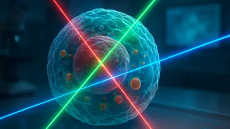
The emergence of protein hydrogels designed through generative AI demonstrates a predictable correlation between calculated binding energies and the physical performance of synthetic materials. This finding serves as a pivotal indicator of how machine learning is evolving from basic pattern

The development of xvr marks a shift toward unified surgical guidance where information from multiple imaging modalities is synthesized into actionable 3D data. Modern medicine has undergone a radical transformation through the rise of minimally invasive procedures, which allow surgeons to perform

Future clinical integration of landmark detection for the carina will allow for fully autonomous safety assessments of endotracheal tube placement in the most vulnerable patients. This technological leap addresses one of the most persistent and high-stakes challenges in pediatric critical care: the

Introduction: The New Standard in Cardiac Triage The rapid evolution of cardiac diagnostics has reached a pivotal juncture where artificial intelligence no longer simply assists physicians but actively redefines the speed and accuracy of life-saving interventions in emergency departments across the
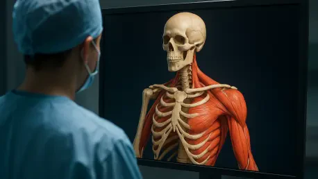
Automating the segmentation of healthy tissues from whole-body scans provides clinicians with a detailed map of a patient's physical resilience and treatment response. For years, the primary objective of whole-body MRI in the oncology ward was to visualize the extent of malignancy and monitor tumor

Advanced generative modeling can now steer patches of a light-microscopy image toward their most plausible high-resolution counterparts using optimal transport theory. This breakthrough addresses the central conflict in modern neurobiology, where researchers must choose between the expansive speed